
|
 |
A Tool For Environmental Science
X-rays are especially valuable for molecular environmental science, because the wavelengths at which a metal atom in a molecule absorbs x-ray photons reveal its oxidation state. The importance of this fact to environmental research is currently being demonstrated in several on-going experiments. In one study, scientists have shown that green rust, a harmless natural oxide of iron common to many soils, can chemically convert a hazardous form of the mineral selenium into a form that is safe to humans and wildlife. Prior to this study, which was led by Satish Myneni and Tetsu Tokunaga, of Berkeley Lab's Earth Sciences Division (ESD), it was thought that only microorganisms could immobilize the selenium contamination that can occur as a result of agricultural runoff. This new knowledge may lead to an alternative way to counteract abnormally high and potentially lethal concentrations of selenium and other trace contaminants in soils and sediments. Myneni and his colleagues are now using x-ray microscopy to analyze samples collected from selenium-contaminated sites for the presence of green rust.
Another ESD researcher, Geraldine Lamble, is using ALS x-rays to learn more about "phytoremediation"—an approach to environmental cleanup which seeks to capitalize on the ability of some plants and fungi to sop up great quantities of soil-borne toxins and convert them into harmless chemical species. Working with a form of x-ray absorption fine structure spectroscopy (XAFS), a technique that can be used to detect and characterize even trace amounts of contaminants in solution, Lamble has been studying a fungi from Norway known to "hyperaccumulate" zinc and other heavy metals which are severely polluting forest soils there. So far, Lamble has established that the zinc is concentrated within the fungal tissue in spots only a few microns in size, and that most of it is being converted into an insoluble chemical compound called zinc oxalate. For cleaning up environments that have been polluted with toxic metals, the best bet remains bioremediation—the use of bacteria and other microorganisms. Lacking the proteins that plants and animals have for storing iron and other essential metals, many of these microorganisms have evolved unique chemical strategies for extracting essential metal ions from their surroundings. Scientists would like to adapt these strategies for the removal of toxic metals from the environment but need a much clearer picture of how they work. Again, ALS x-rays can help, as evidenced by Brian Tonner, from the University of Central Florida, who used the spectromicroscopy and x-ray microscopy beamlines to obtain detailed micrographs of bacteria that are known to absorb manganese from water and oxidize it into manganite, an insoluble mineral. David Shuh, a chemist with Berkeley Lab's Chemical Sciences Division, uses ALS x-rays to study actinides, the group of radioactive elements that have been linked to various types of cancers, and whose presence complicates any waste cleanup or storage effort. Explains Shuh, "If you want to sequester the actinides from other wastes, you need to understand complex actinide chemistry, things that people couldn't get at directly before the advent of synchrotron radiation studies." The ALS is well-suited for studying atmospheric chemistry as well, but for this research, the preferred photons are at the vacuum ultraviolet (VUV) energies. Working at undulator Beamline 9.0.2.2, Cheuk-Yiu Ng, a chemist who holds a joint appointment with the Ames Laboratory and Iowa State University, led a team that used VUV spectroscopy to take a new look at oxygen. About 20-percent of the earth's atmosphere consists of oxygen molecules which help shield the planet's surface from lethal doses of ultraviolet radiation. Understanding how oxygen absorbs uv rays and the subsequent chemistry that leads to the regeneration of more oxygen is of vital concern to atmospheric chemists. When an oxygen molecule absorbs a VUV photon, it ejects an electron and becomes a cation—a positively charged ion. This process is called photoionization. The ultimate fate of an oxygen molecule is determined by its electronic structure after photoionization takes place. Ng and his colleagues used VUV light from the ALS and a technique called "pulsed-field ionization photoelectron spectroscopy" to obtain the most detailed picture ever recorded of an oxygen cation's electronic structure. This approach should prove equally applicable to other atmospheric molecules including nitrogen and ozone. "We succeeded by adapting a laser spectroscopy technique to synchrotron radiation," says Ng. "The ALS with its bright beams of VUV light over a wide range of wavelengths made this adaptation possible." Although the ALS is designed for peak production of soft x-ray and VUV light, it still produces photons below those energies on the spectrum. For example, Beamline 1.4, which operates off a bend-magnet and serves three experimental endstations, generates beams of infrared (IR) light at far higher brightness than conventional IR sources such as Globars (TM), which are silicon-carbide filaments that radiate IR light when heated. The ability to focus IR light down to a 10 micron spot size opens up new possibilities for environmental studies and other areas of research. A molecule, because of the non-stop motion of its atoms, vibrates at a characteristic
frequency within the IR spectrum that can be used to identify its type, much like a
fingerprint can identify a person. When a molecule is struck by an IR photon that matches
its vibrational frequency, it will resonate. This resonance can be detected through a
variety of spectroscopic techniques and used to not only "fingerprint" the
molecule, but to also measure and manipulate it.
Michael Martin, manager of Beamline 1.4 says, "We can shine our (IR) beam on an unknown sample and determine its chemical composition. With our fast time resolutions (a spectral analysis can be recorded every five nanoseconds), we can then observe changes in this chemical composition while they are taking place and see which molecular species go away as they turn into something else." Although the bend magnet for beamline 1.4 probably has the poorest focus in the ALS for XUV research, Martin says it is "just fine" for IR spectroscopy. As a result, ALS light that might otherwise have been discarded is being put to good use by research teams such as the one led by Donald Sparks, who chairs the University of Delaware's Department of Plant and Soil Sciences. Sparks and his team are using IR photons to do molecular-scale studies of reactions between chemicals that occur naturally in a soil and those that are introduced by human activity such as metals, industrial chemicals, and pesticides. In addition to environmental studies, IR photons at the ALS are also being used in studies aimed at discovering new types of materials; measuring energy bandgaps in novel semiconductors and high-Tc superconductors; investigating sub-monolayers of atoms adsorbed onto surfaces during interface reactions and corrosion; and probing the reactive transients, the molecules that form temporarily during the intermediate stages of a chemical reaction and often actually determine the reaction's outcome. Martin, who is a solid-state physicist, has worked with Wayne McKinney, the principle designer of Beamline 1.4, to ensure that the IR experimental endstations are second to none in user friendliness. "The goal is for users to be able to come in, click a button, and get their data without having to understand how the beamline system works," Martin says. "This is how you reach out to users who want to come here to do science and not to learn beam physics." |
||||||
|
|||||||
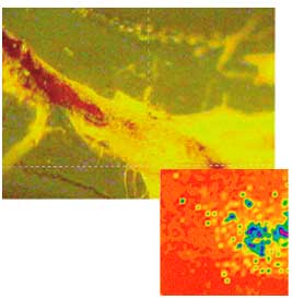 Certain fungi in Norwegian forest
soils have been found to "hyperaccumulate" zinc and other heavy metal
pollutants. A fluorescence map shows the zinc to be concentrated within the fungal tissue
in spots only a few microns in size.
Certain fungi in Norwegian forest
soils have been found to "hyperaccumulate" zinc and other heavy metal
pollutants. A fluorescence map shows the zinc to be concentrated within the fungal tissue
in spots only a few microns in size. 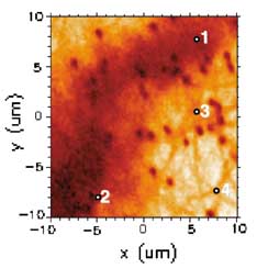 Illumination with ALS x-rays reveals
microscale chemical heterogeneity in organic soil samples that could play a role in
macroscale remediation efforts.
Illumination with ALS x-rays reveals
microscale chemical heterogeneity in organic soil samples that could play a role in
macroscale remediation efforts. 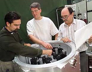 Working with infrared microscopy at an
ALS experimental station, physicist Michael Martin (center), manager of Beamline 1.4,
helped two Bay Area science teachers develop a classroom study unit as part of an
education partnership program held at Berkeley Lab.
Working with infrared microscopy at an
ALS experimental station, physicist Michael Martin (center), manager of Beamline 1.4,
helped two Bay Area science teachers develop a classroom study unit as part of an
education partnership program held at Berkeley Lab.