
|
 |
Insights In Biosciences and Health
One of the busiest ALS beamlines is Beamline 5.0 which serves the Macromolecular Crystallography Facility (MCF). Sending a beam of x-rays through a crystal creates a diffraction pattern when the photons are scattered by the crystal's atoms. This pattern can be translated by computer into 3-D images of the crystal. Identifying the crystal structure of a protein or other type of biological molecule is critical to a wide variety of pursuits including rational drug-design for safer and more effective treatment of diseases and disorders. Powered by a 38-pole wiggler magnet, Beamline 5.0 provides photons of x-rays ranging in wavelengths from 0.9 to 4.0 angstroms and in energies from 3.5 to 14 keV (thousand electron volts). Increased energy means increased penetration which, coupled with precise tuning, makes these photons ideal for protein crystallography. Many of its users consider it to be one of the world's best. "One of our strengths is that we run a total scientific program here," says
Thomas Earnest, a biophysicist with Berkeley Lab's Physical Biosciences Division who now
oversees activity on the MCF after having been in charge of its design and construction.
As the head of a research group investigating proteins in cell membranes, Earnest
understands crystallographer needs. Rather than constructing beamlines other researchers
might want to use, he says, "We build beamlines on which we ourselves want to do
science." This intimate knowledge of the needs of its users, has helped make the MCF
one of the most user-friendly of all the ALS experimental stations. Thanks to taking full
advantage of a technique called MAD (multiple-wavelength anomalous diffraction), which
greatly speeds up the process of crystallography data collection, the MCF is also one of
fastest stations for getting results.
A major aim of protein crystallography is rational drug-design, the strategy in which a pharmaceutical drug is targeted to inhibit or promote the activity of a specific protein. Scientists from Roche Biosciences have already used data obtained at the MCF to solve the crystal structures of more than a dozen protein-inhibitor complexes as part of their effort to design better treatments for inflammatory diseases. The intensity and focusability of the photons at the MCF enabled the Roche scientists to work with tiny frozen microcrystals measuring no more than a few hundredths of a millimeter in diameter. Cancer research is also benefiting from MCF x-rays. Researchers led by Barry Stoddard from the Fred Hutchinson Cancer Research Center in Seattle, an affiliate of the National Cancer Institute, are studying how segments of DNA and cell proteins called histones combine and fold into fibers of chromatin and how this arrangement can be erroneously altered. This could be an indicator of genetic susceptibility to cancerous mutations. At the MCF, Stoddard and his collaborators have already identified the crystal structures of a pair of intron-encoded restriction enzymes, proteins that segment DNA at specific sites along the molecule. In what could prove to be historic research, a group led by Harry Noller, a molecular biology professor at UC Santa Cruz, is gathering data on the crystal structure of a ribosome. In solving this structure, they hope to solve the mystery of ribosomal RNA. Ribosomes are tiny cell organelles responsible for protein synthesis. They receive and somehow put together "messenger RNA" molecules from the nucleus, which carry the genetic code for assembling proteins, and "transfer RNA" molecules from the cytoplasm which carry the amino acids from which proteins are made. No one knows exactly how this process works, but it is known that a major component of ribosomes is a third type of RNA. Determining the structure of ribosomes would be a giant step toward understanding the mechanism by which this critical organelle functions, but the job is difficult. Though smaller than most viruses, a ribosome is a very large molecular complex, consisting of three RNA and more than 50 protein molecules. Says Earnest, "Obtaining atomic-resolution data for so large a macromolecular
complex can only be done on a high-brightness synchrotron source such as the ALS."
While protein crystallography is a well-established research tool, x-ray microscopy must still prove itself. There are those in the biosciences who question whether x-ray microscopy can bring anything new to their field. Even hard-core skeptics should be convinced by what has already been accomplished at Beamline 6.1.2, also known as XM-1. Drawing light from a bend magnet, XM-1 is a direct-imaging transmission x-ray microscope whose photon-energy range extends from 250 to 950 electron volts. Within this range falls the so-called "water window," the energy span over which x-rays don't "see" water, but do "see" carbon-containing materials. This means that high-contrast images of proteins and other interior cell structures can be obtained at XM-1 without the need for staining. Werner Meyer-Ilse, a microscopist with Berkeley Lab's Center for X-Ray Optics, directed the design and construction of XM-1 and now oversees the research conducted with it. Like Earnest, he, too, understands the needs of his beamline's users. "My personal interest is to develop x-ray microscopy and I am only able to do this in a collaborative manner," he says. "Therefore all of the users of this beamline, from all their different scientific fields, become my collaborators in developing the technology." Light is being shed by XM-1 photons on a number of long-standing biological mysteries. For example, high-magnification images are being obtained of the malaria parasite when it is inside a red blood cell, providing new information on one of the oldest and most persistent of all human diseases. Revealing images are also being obtained of the relationship between breast cell nuclei in response to a mass of fibrous and globular proteins outside of the cells called the Extracellular Matrix or ECM. Research over the past two decades by Mina Bissell, director of Berkeley Lab's Life Sciences Division, has shown that the "dialogue" between breast cells and the ECM is important for normal function. Bissell and one of her colleagues, Sophie Lelievre, are now using x-ray microscopy to study the signaling mechanism entailed in this dialogue and how it may go wrong in malignancy. A recent development at XM-1 may help resolve the debate over whether or not a cell nucleus has an organized internal structure. Meyer-Ilse and Carolyn Larabell, a Berkeley Lab Life Sciences Division cell biologist/microscopist, have obtained novel x-ray microscopy images of labeled proteins in breast cell nuclei which have been fixed, but were intact and in a hydrated state. These images showed a resolution between 30 and 50 nanometers compared to the 200 nanometer resolution of images obtained with the best visible light microscopes. "Soft x-ray microscopy is now poised to make a major contribution to the understanding of cell function and structure," Larabell says. "We have demonstrated a practical technique that gives cell biologists a whole new way of looking at their samples." Sample preparation is critical to the success of this new technique. Antibodies, bound to nano-sized gold particles, are tagged to specific proteins within the cell, including some inside the nucleus. Because the gold particles are too small to be seen even with x-rays, the proteins are further enhanced with a coating of silver, similar to the use of silver emulsions to bring out images on photographic film. Subsequent exposure to x-rays provides sharp images of these labeled proteins. The combination of gentle cell fixation, protein-labeling and the unique capabilities of XM-1 is a winner. Already, it has been used to obtain dramatic images of tubulin, an important protein of the cell's skeletal network. This proof-of-principle experiment is merely a preview of coming attractions at XM-1. |
||||||
|
|||||||
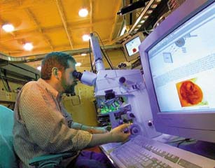 Werner Meyer-Ilse oversees research
at XM-1, a direct-imaging transmission x-ray microscope that is being used in a wide range
of studies, including the malaria parasite when it is inside a red blood cell (pictured on
screen).
Werner Meyer-Ilse oversees research
at XM-1, a direct-imaging transmission x-ray microscope that is being used in a wide range
of studies, including the malaria parasite when it is inside a red blood cell (pictured on
screen). 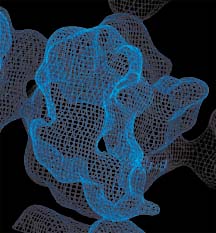 Protein
crystallography at the ALS yielded this electron density map of a ribosome, a cell
organelle consisting of three RNA and more than 50 protein molecules that is responsible
for protein synthesis.
Protein
crystallography at the ALS yielded this electron density map of a ribosome, a cell
organelle consisting of three RNA and more than 50 protein molecules that is responsible
for protein synthesis. 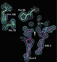 X-ray beams at
the Macromolecular Crystallography Facility helped researchers from the Fred Hutchinson
Cancer Research Center solve the crystal structure of Ppo-I, a restriction enzyme that
cuts DNA at specific sites along the molecule.
X-ray beams at
the Macromolecular Crystallography Facility helped researchers from the Fred Hutchinson
Cancer Research Center solve the crystal structure of Ppo-I, a restriction enzyme that
cuts DNA at specific sites along the molecule.