Introduction
Over
the past few decades, there has been much interest in the
structure
and properties of mineralized biological tissues like bone and dentin
(a
structurally simpler analogue of bone that makes up the bulk of the
human
tooth); in particular, considerable research have been focused on their
mechanical properties and into how they fracture. An
understanding
of these properties is of great importance from the perspective of
developing
a realistic framework for life prediction, particularly in light of the
effect that microstructural modifications from aging, disease,
remodeling,
etc., can have in degrading the tissue. Central to these issues
is
the fracture toughness of these materials, which characterizes their
resistance
to incipient cracking and fracture, and the microstructural mechanisms
that are the source of such resistance. Understanding such
properties
in the context of the inherent hierarchical complexity of the
microstructure
of these tissues (Fig. 1) is of obvious importance. However,
surprisingly,
such questions have largely remained unanswered and to a large extent,
even uninvestigated.
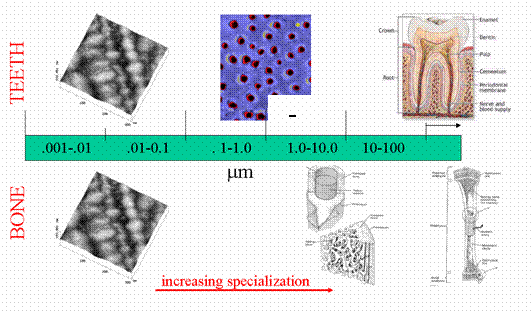
Fig.
1: Hierarchy of
the microstructure
of two common mineralized tissues, human teeth and bone are shown
here.
Though very different at higher scales of organization, fundamentally,
both tissues are comprised of collagen fibers.
Evidence based on microscopic observations has indicated that a number of mechanisms that could “toughen” mineralized tissues are active. Indeed, it has been observed that microcracks and crack bridges form during the fracture of both materials. Typically microcracks preferentially form at the peritubular cuffs within the inelastic zone surrounding a macroscopic crack in (particularly human) dentin and around osteons, due to osteon-matrix interface debonding or osteon pull-out, in bone. Crack bridging, conversely, have been suggested to occur by uncracked ligaments and/or collagen fibers in both dentin and bone. Fig. 2 shows some typical examples of such mechanisms.
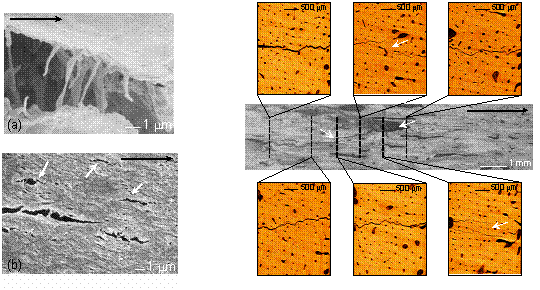
Fig. 2:
Scanning electron micrographs of (a) crack bridging by collagen fibers
in human cortical bone, and (b) microcracking (indicated by white
arrows)
at tubule sites in dentin. (c) Evidence of uncracked-ligament bridging
(indicated by white arrows) shown for a crack in 61-year old female
cortical
bone in an optical micrograph (center) and in x-ray computed
tomographic
reconstructions of through-thickness slices. The horizontal black
arrow in each case indicates the direction of nominal crack
growth.
(Tomography was performed at the Stanford Synchrotron Radiation
Laboratory,
SSRL, Menlo Park, CA)
Although, there are differences in the architecture of various
mineralized
tissues, at the nano-scale, they are fundamentally quite similar with a
network (almost matte) of collagen fibers forming the basis of the
microstructure.
Consequently, this study aims at furthering our understanding of the
macroscopic
fracture behavior in the context of the underlying collagen-based
nano-structure.
Such research is believed to be critical to the development of a
micromechanical
fracture mechanics based framework for understanding the problem of
fracture
and fatigue failure in mineralized tissue.
Current Researchers
J. H. Kinney
Recent
Publications
![]() R.
K. Nalla, J. J. Kruzic, J. H. Kinney, R. O. Ritchie, "Mechanistic
Aspects of Fracture and R-Curve Behavior in Human Cortical Bone ", Biomaterials, 2004.
R.
K. Nalla, J. J. Kruzic, J. H. Kinney, R. O. Ritchie, "Mechanistic
Aspects of Fracture and R-Curve Behavior in Human Cortical Bone ", Biomaterials, 2004.
![]() V.
Imbeni, R.K. Nalla, C. Bosi, J.H. Kinney, and R.O. Ritchie, “On
the in vitro fracture toughness of human dentin”, J. Biomed. Mater.
Res., 2003; 66A:1-9.
V.
Imbeni, R.K. Nalla, C. Bosi, J.H. Kinney, and R.O. Ritchie, “On
the in vitro fracture toughness of human dentin”, J. Biomed. Mater.
Res., 2003; 66A:1-9.
![]() R.K.
Nalla, V. Imbeni, J.H. Kinney, M. Staninec, S.J. Marshall and R.O.
Ritchie,
“On
the in vitro fatigue behavior of human dentin with implications for
life
prediction”, J. Biomed. Mater. Res., 2003; 66A:10-20.
R.K.
Nalla, V. Imbeni, J.H. Kinney, M. Staninec, S.J. Marshall and R.O.
Ritchie,
“On
the in vitro fatigue behavior of human dentin with implications for
life
prediction”, J. Biomed. Mater. Res., 2003; 66A:10-20.
![]() R.K.
Nalla, J.H. Kinney, and R.O. Ritchie, “On
the fracture of human dentin- Is it stress- or strain-controlled?”,
J. Biomed. Mater. Res., 2003. 67A: 484–495.
R.K.
Nalla, J.H. Kinney, and R.O. Ritchie, “On
the fracture of human dentin- Is it stress- or strain-controlled?”,
J. Biomed. Mater. Res., 2003. 67A: 484–495.
![]() R.K.
Nalla, J.H. Kinney, and R.O. Ritchie, “Effect
of orientation on the in vitro fracture toughness of dentin: The role
of
toughening mechanisms”, Biomater., 2003; 24:3955-3968.
R.K.
Nalla, J.H. Kinney, and R.O. Ritchie, “Effect
of orientation on the in vitro fracture toughness of dentin: The role
of
toughening mechanisms”, Biomater., 2003; 24:3955-3968.
![]() R.K.
Nalla, J.H. Kinney, and R.O. Ritchie, “Mechanistic
Fracture Criteria for the Failure of Human Cortical Bone”, Nature
Mater.,
2003; 2:164-168.
R.K.
Nalla, J.H. Kinney, and R.O. Ritchie, “Mechanistic
Fracture Criteria for the Failure of Human Cortical Bone”, Nature
Mater.,
2003; 2:164-168.
![]() R.K.
Nalla, J.H. Kinney, S.J. Marshall and R.O. Ritchie, “On
the In Vitro Fatigue Behavior of Human Dentin: Effect of Mean Stress”,
J. Dent. Res., 2004; 83(3):211-215.
R.K.
Nalla, J.H. Kinney, S.J. Marshall and R.O. Ritchie, “On
the In Vitro Fatigue Behavior of Human Dentin: Effect of Mean Stress”,
J. Dent. Res., 2004; 83(3):211-215.
![]() J.J.
Kruzic, R.K. Nalla, J.H. Kinney and R.O. Ritchie, “Crack
blunting, crack bridging and resistance-curve fracture mechanics in
dentin:
Effect of hydration”, Biomater., 2003; 24:5209-5221.
J.J.
Kruzic, R.K. Nalla, J.H. Kinney and R.O. Ritchie, “Crack
blunting, crack bridging and resistance-curve fracture mechanics in
dentin:
Effect of hydration”, Biomater., 2003; 24:5209-5221.
![]() V.
Imbeni, R.K. Nalla, J.H. Kinney, M. Staninec, S.J. Marshall, G.W.
Marshall
and R.O. Ritchie, “Stress/Life Cyclic Fatigue Behavior of Human
Dentin”,
in: Proc. IADR/AADR/CADR meeting (J. Dent. Res.), San Diego, CA, March
2002. [View
poster]
V.
Imbeni, R.K. Nalla, J.H. Kinney, M. Staninec, S.J. Marshall, G.W.
Marshall
and R.O. Ritchie, “Stress/Life Cyclic Fatigue Behavior of Human
Dentin”,
in: Proc. IADR/AADR/CADR meeting (J. Dent. Res.), San Diego, CA, March
2002. [View
poster]
![]() R.K.
Nalla, V. Imbeni, J.H. Kinney, S.J. Marshall and R.O. Ritchie, “On the
development of life prediction methodologies for the failure of human
teeth”,
submitted to: Proc. Symposium on Materials Lifetime Science and
Engineering,
Eds.: P.K. Liaw et al.), TMS, San Diego, CA, March 2003. [View
paper] [View
presentation]
R.K.
Nalla, V. Imbeni, J.H. Kinney, S.J. Marshall and R.O. Ritchie, “On the
development of life prediction methodologies for the failure of human
teeth”,
submitted to: Proc. Symposium on Materials Lifetime Science and
Engineering,
Eds.: P.K. Liaw et al.), TMS, San Diego, CA, March 2003. [View
paper] [View
presentation]
![]() R.K.
Nalla, V. Imbeni, J.H. Kinney, M. Staninec, S.J. Marshall, G.W.
Marshall
and R.O. Ritchie, “On the Development of a Fracture Mechanics-Based
Approach
to Failure Prediction in Human Dentin”, in: Proc. IADR/AADR/CADR
meeting,
San Antonio, TX, March 2003. [View
poster]
R.K.
Nalla, V. Imbeni, J.H. Kinney, M. Staninec, S.J. Marshall, G.W.
Marshall
and R.O. Ritchie, “On the Development of a Fracture Mechanics-Based
Approach
to Failure Prediction in Human Dentin”, in: Proc. IADR/AADR/CADR
meeting,
San Antonio, TX, March 2003. [View
poster]
![]() R.
O. Ritchie, C. L. Muhlstein, and R. K. Nalla, “On the Fatigue and
Fracture
of 'Nano' and 'Bio' Materials”, in: Proceedings of the International
Conference
on Advanced Technology in Experimental Mechanics 2003 (ATEM'03), H.
Kimura,
ed., 2003.
R.
O. Ritchie, C. L. Muhlstein, and R. K. Nalla, “On the Fatigue and
Fracture
of 'Nano' and 'Bio' Materials”, in: Proceedings of the International
Conference
on Advanced Technology in Experimental Mechanics 2003 (ATEM'03), H.
Kimura,
ed., 2003.
![]() A.E. Porter, R.K. Nalla, J.H. Kinney
and R.O. Ritchie, “Changes in mineralization with aging-induced
transparency in human root dentin”, in: The 2004 Gordon Research
Conference on Biomineralization, New London, NH, August 2004. [View
Poster]
A.E. Porter, R.K. Nalla, J.H. Kinney
and R.O. Ritchie, “Changes in mineralization with aging-induced
transparency in human root dentin”, in: The 2004 Gordon Research
Conference on Biomineralization, New London, NH, August 2004. [View
Poster]