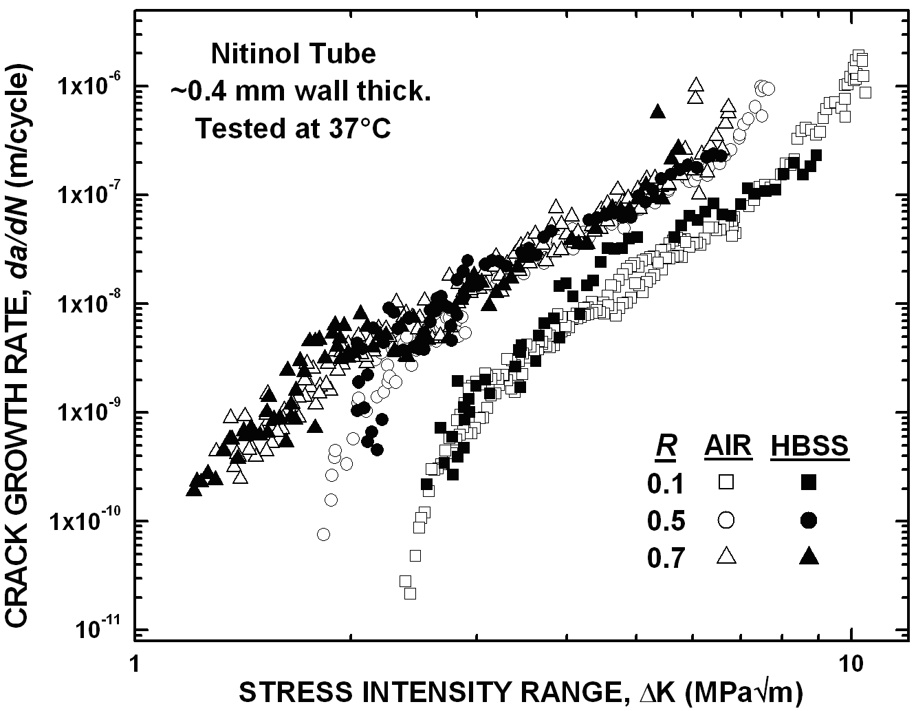
Ritchie Group Research Program in Nitinol

Research Program on the Fatigue and Fracture of Superelastic Nitinol

Program Description

Biomedical devices manufactured from Nitinol tube (e.g. Johnson & Johnson's S.M.A.R.T. Stent) invariably experience in vivo loading conditions that cause a variety of fatigue and overload conditions. These loading scenarios can, in the best case scenario lead to minor fracture in the device with no negative consequences to the device longevity, and in the worse case complete failure of the product potentially resulting in harm to the patient. Consequently, it is the primary objective of the current work to provide a comprehensive characterization of the in vitro fatigue-crack growth properties (especially at the all important near-threshold growth rates) and fracture toughness behavior in thin-walled Nitinol tubing typically used in commercial stent manufacture in simulated physiological environment, in order to realize quantifiable engineering parameters for designing against premature failure from overload and/or in vivo fatigue damage in endovascular self-expanding stents.

This work combines standard fracture mechanics test techniques (e.g. fatigue crack-growth curve generation, R-curve testing, etc), X-ray diffraction for texture analysis, and high-spatial-resolution X-ray Microdiffraction at the Advanced Light Source Beamline 7.3.3 at the Lawrence Berkeley National Laboratory. By combining these test techniques, we have learned about the fracture behavior, fatigue characteristics, and the roles of texture and phase transformation on the crack-growth properties of Nitinol tube.


Figure 1: Fatigue crack-growth behavior of flattened Nitinol tube evaluated in 37°C air and Hanks' balanced saline solution (HBSS) simulated body fluid, showing a dependence on the load ratio, R.


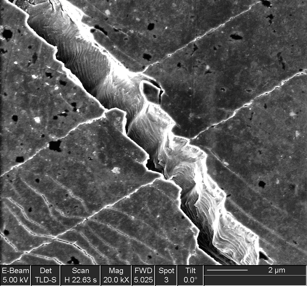
Figure 2: Evidence of the effects of texture in Nitinol manifesting as a fatigue crack angled to the pure Mode I crack path (left to right crack advance).


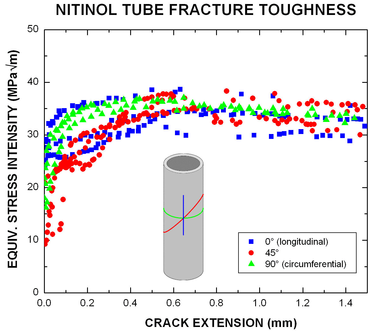
Figure 3: R-curve behavior in Nitinol tube showing fracture toughness dependence on the crack angle with respect to the drawing direction (lowest initiation toughness in the 45° direction).


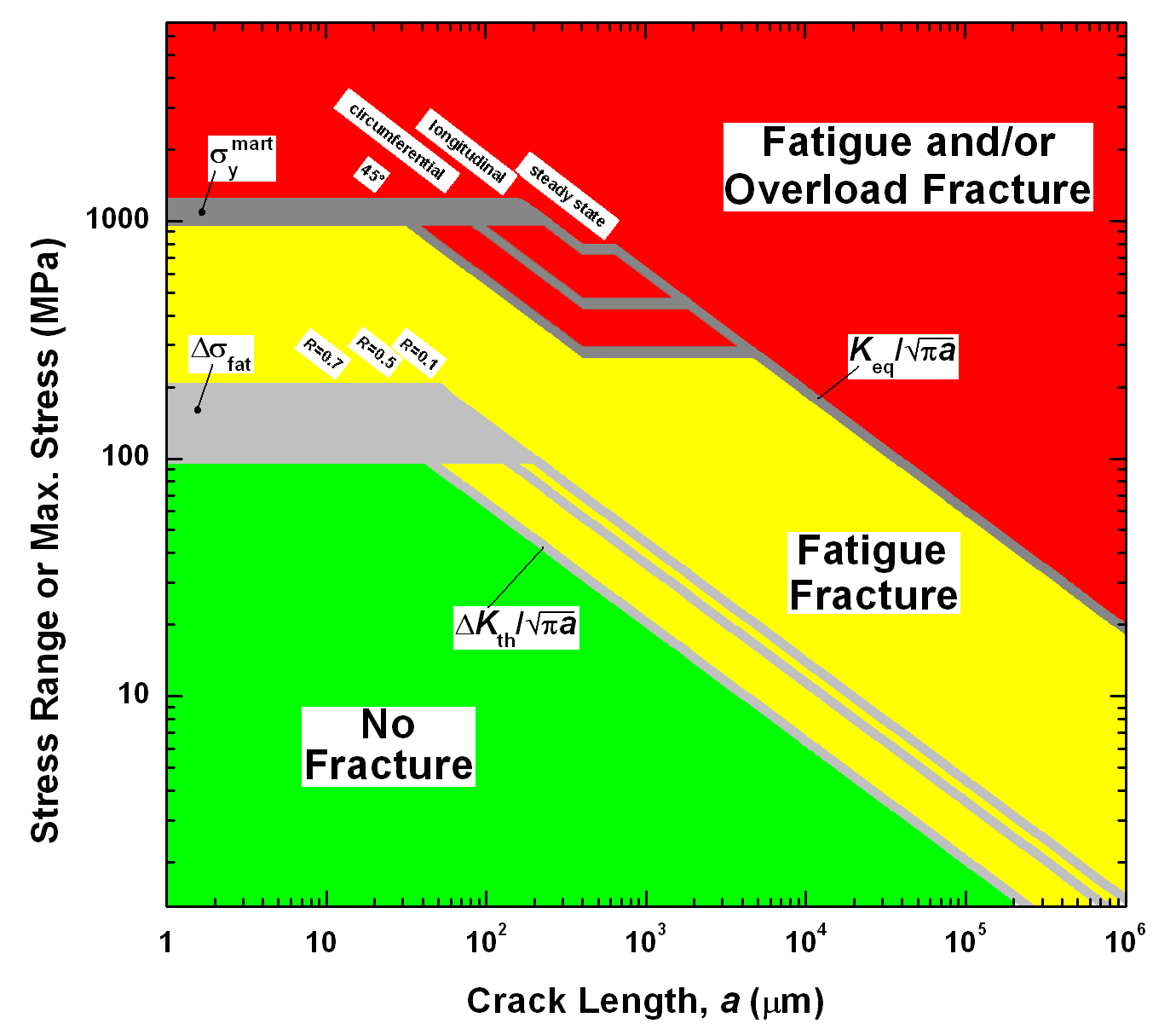
Figure 4: Kitagawa-Tagahashi safe operating zone plot for fracture and fatigue conditions in Nitinol tube. Note a generic calculation for stress intensity was used, such that the geometry factor, Y, was set to unity in the relation K = Y a; exact stress intensity calculations need to be performed to accurately adapt this plot to a real biomedical device geometry.


Figure 5: Evolution of a fatigue-induced transformation zone ahead of an atomically sharp crack as determined by X-ray Microdiffraction. Notice the grain-dependant transformation showing suppression of the transformation in grains oriented near the <100> direction.


Online Publications


Ritchie Group
LBNL , MSD
Dept of MSME , UC Berkeley

Last updated 08/03