Mixed-Mode
Fracture of Human
Cortical Bone
Fracture
studies on the
behavior of human cortical bone have provided much information on how
the
hierarchical microstructure of bone (see Fig. 1a) is able to resist the
initiation and growth of incipient cracks at numerous length
scales. Of
particular importance is how the nano/microstructure
can affect the crack path, which in turn controls the specific fracture
resistance. The structure of bone is highly anisotropic with a
preferred microstructural crack path
aligned along the long axis of
the bone in the form of the osteonal
cement lines,
the highly mineralized interfaces between the lamellae and the Haversian canals. As fractures in the
transverse
(breaking) orientation (see Fig. 1b) are nominally aligned
perpendicular to
this weaker direction (i.e., parallel to the osteons),
the toughness of human cortical bone is far higher in the
transverse, as
compared to the longitudinal, orientation -- it is easier to split than
to
break.
However,
to date most
such studies on the fracture toughness of bone have been performed
under
tensile (mode I) loading, the underlying assumption being that the mode
I
toughness value is the lowest (as is the case for most
materials).
However, such loading conditions are not typical of those experienced
physiologically; moreover, due to the marked anisotropy of the
bone-matrix
structure, mode I loading is not necessarily worst-case.
Accordingly,
we are
examining the fracture mechanics of human cortical bone under
mixed-mode loads,
specifically under mode I (tensile opening), mode II (in-plane shear)
and mode
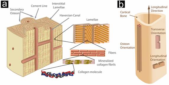
Figure 1. (a) Bone is a composite of
collagen
molecules (~1.5 nm in diameter) and hydroxyapatite
crystals (HA). The HA is deposited between the heads and tails of the
collagen
molecules, which are in a staggered array; this structure is called a
mineralized collagen fibril. The mineralized collagen fibrils form
arrays
called fibers and the fibers assemble into arrays called lamellae (~5-mm
thick), which resemble
sheets. The
lamellae are concentrically arranged around a central vascular channel.
This
whole structure is called a secondary osteon
(~250
mm
in diameter) and is
aligned
parallel to the longitudinal axis of the bone. The lamellae in the
secondary osteon are separated from the
interstitial lamellae by a hypermineralized
layer of material called a cement line
(~5-mm thick).
(b) Due to the anisotropic nature of this structure, the toughness must
be
assessed in two different orientations. In the longitudinal
orientation, the
crack is parallel to the orientation of the osteons
while in the transverse orientation, the crack is perpendicular to the
orientation of the osteons.

Figure 2. In fracture mechanics, the
stress
intensity due to any loading scenario can be broken down into three
modes of
loading: (a) tensile opening -- mode I, (b) in-plane shear -- mode II,
and (c)
anti-plane shear -- mode
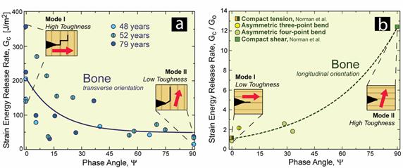
Figure 3. (a) For human cortical bone
oriented in
the transverse orientation, the toughness (measured in terms of the
critical
strain energy release rate) is higher in mode I (tension) than mode II
(in-plane shear). In this orientation, the preferred microstructural
path (dark brown lines in schematic) is perpendicular to the
orientation of the
crack. When a mode II driving force is applied, the direction of the
preferred
mechanical path is at a 74° angle to the original plane of the crack
(pink
arrow in schematic); thus, the preferred direction of the
microstructure and
the driving force are commensurate and bone has a low toughness. For
mode I,
the direction of the driving force and the preferred microstructural
path are at a 90° angle, which results in a high toughness. (b)
The
opposite relation occurs in the longitudinal orientation, where the
crack is
oriented parallel to the preferred microstructural
path. In this case, bone is tougher in shear than tension. This figure
is
supplemented with data from Norman et al.
[2].
Current
Researchers
References
[1]
Zimmermann EA, Launey ME, Barth
HD, Ritchie RO. Biomaterials. 2009;30(29):5877-84.
[2] Norman
TL, Nivargikar SV, Burr DB. J Biomech. 1996;29(8):1023-31.