One major use of radioisotopes is in nuclear medicine. Of the 30 million people who are hospitalized each year in the United States, 1/3 are treated with nuclear medicine. More than 10 million nuclear-medicine procedures are performed on patients and more than 100 million nuclear-medicine tests are performed each year in the United States alone. A comparable number of such procedures are performed in the rest of the world.
There are nearly one hundred radioisotopes whose beta and/or gamma radiation is used in diagnosis, therapy, or investigations in nuclear medicine. The most used radioisotopes were discovered before World War II using the early cyclotrons of Ernest Lawrence, with the initial applications to medicine being developed by his brother John Lawrence. Some of the most well known radioisotopes, discovered by Glenn Seaborg and his coworkers, are 131I (discovered in 1938), 60Co (1937), 99mTc (1938), and 137Cs (1941). By 1970, 90 percent of the 8 million administrations per year of radioisotopes in the United States utilized either 131I, 60Co, or 99mTc. Today, 99mTc, with a half-life of 6 hours, is the workhorse of nuclear medicine. It accounts for more than 10 million diagnostic procedures a year in the United States. It is used for brain, bone, liver, spleen, kidney, lung and thyroid imaging as well as for blood-flow studies.
131I, with a half-life of 8 days, is used to diagnose and treat thyroid disorders. Seaborg’s mother was one of the first to benefit from the use of this radioisotope that her son had discovered. Fatally ill from hyperthyroidism, (a related condition from which her sister died), diagnosis and treatment with 131I led to her complete recovery and a long life. Former President George Bush and First Lady Barbara Bush are some notable people who were successfully treated for Graves' disease, a thyroid disease, with 131I. Radioactive iodine treatment is so successful that it has virtually replaced thyroid surgery.
A very effective role for radioisotopes in nuclear medicine is the use of short-lived positron emitters such as 11C, 13N, 15O, or 18F in a process known as Positron Emission Tomography (PET). Incorporated in chemical compounds that selectively migrate to specific organs in the body, diagnosis is effected by detecting annihilation gamma rays–two gamma rays of identical energy emitted when a positron and an electron annihilate each other. These gamma rays have the very useful property that they are emitted in exactly opposite directions. When both are detected, a computer system may be used to reconstruct where the annihilation occurred. By attaching a positron emitter to a protein or a glucose molecule, and allowing the body to metabolize it, we can study the functional aspect of an organ such as the human brain. The PET image shows where the glucose has been absorbed (Fig. 13-3a).
PET imaging becomes even more valuable when we can observe the functional image compared to the anatomical image. Magnetic Resonance Imaging (MRI)–originally known as Nuclear Magnetic Resonance Imaging–can provide very detailed images of the anatomy as shown in the second image shown in Fig. 13-3b. These techniques provide researchers a better understanding of what is healthy tissue versus what is diseased.
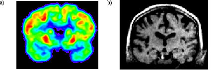 Fig. 13-3. a) PET scan image of the human brain. b) MRI image of the human brain.