his past year, Berkeley Lab biophysicists, led by Eva Nogales and Kenneth Downing, along with Sharon Wolf, put the finishing touches on a 3-dimensional atomic model of tubulin that has been six years in the making. At a resolution of 3.7 angstroms, the model provides the first highly-detailed look at one of Nature's most essential proteins.
Tubulin is a "heterodimer," meaning it consists of a pair of polypeptide chains-called monomers-that differ in the sequence of their amino acids. The protein's importance stems from its flexibility. Polypeptide chains of tubulin curl around to form tiny hollow fibers called microtubules that intermesh to provide a cell with its cytoskeletal framework. Thanks to the flexibility of the tubulin protein, microtubules can shift through a variety of different formations. This formation-shifting is what enables a cell to carry out such vital activities as mitosis (cell division) and the regulation of materials passing through its membranes.
|
(Click on image to enlarge)
|
|
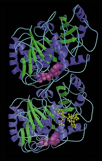
A ribbon diagram based on the 3.7 angstrom resolution model of tubulin developed at Berkeley Lab, shows a dimer consisting of two almost identical monomers. Each is formed by a core of two beta sheets (blue and green) surounded by helices, and each binds to a guanine nucleotide (pink). In addition to a nucleotide binding site, each monomer also has two other binding sites, one for proteins, and one for the anti-cancer drug taxol.
|
The tubulin model created by the Berkeley Lab researchers shows that each of its monomers-the alpha and the beta-is a compact molecular structure with three functional components or domains, one that binds to nucleotides, one that binds to drugs, and one that looks to be a binding site for other proteins.
"The interaction between domains is very tight so that the effects that nucleotides, drugs, and other proteins have on tubulin are firmly linked," says Nogales. "The assembly of tubulin and its regulation through dynamic instability results from the fine tuning of these three components."
Identifying the atomic structure of tubulin's drug binding sites should prove especially valuable for cancer researchers. Taxol, a natural substance found in the bark of the Pacific yew tree, has been shown in clinical tests to be an effective treatment for a number of malignancies, including ovarian, breast, and lung. Cancer occurs when cell division runs amok. By binding to tubulin and causing the protein to lose its flexibility, taxol can prevent cell division from taking place. With better knowledge of the tubulin drug binding site and its interaction with taxol, scientists should be able to synthesize an even more effective anti-cancer drug, one that interacts only with the tubulin of cancerous cells.
To produce their model, the Berkeley Lab researchers first polymerized tubulin proteins under the same conditions in which microtubules are formed, except for the additional presence of zinc. The zinc prevents tubulin chains from curling around into hollow fibers. Instead, the polymerized tubulin forms two-dimensional crystalline sheets that are ideal for imaging by electron crystallography. The use of electron-based rather than the x-ray-based crystallography techniques customarily employed in protein studies was crucial to the model's 3.7 angstrom resolution.
| Lead scientists on this project
|
|
|
|
|
|
"Conventional x-ray crystallography techniques require much more protein than electron crystallography," says Downing. "Obtaining diffraction patterns with an electron beam enables us to work with crystals only one molecule in thickness, giving us our high resolution."
The final 3-D model is a computerized reconstruction derived from a data set that included 93 electron diffraction patterns and 159 images culled from more than 4,000 images. The images were produced on a special electron microscope equipped to allow the crystals to be studied from a variety of different angles with minimum damage.
"We needed to keep our samples extremely stable during the imaging process in order to get the resolution we wanted," says Nogales. To this end, she and her colleagues used taxol to lock their samples into fixed positions for imaging. That the taxol would bind to their crystals is in part a confirmation that their structural model of tubulin is accurate.
- Lynn Yarris