n two separate studies, research teams led by chemist Alexander Pines demonstrated that xenon gas, a common anesthetic, can be specially treated to amplify the volume of MRI signals from any atomic nuclei with which it makes contact. In the first study, Pines and collaborators were able to nearly double the strength of MRI signals from hydrogen nuclei in solution, and to increase the signals of the surface atoms of a solid by a factor of one hundred. In the second study, Pines and Thomas Budinger, head of Berkeley Lab's Center for Functional Imaging, used pre-treated xenon to record the first observations of an anesthetic entering human red blood cells.
MRI, like its cousin, Nuclear Magnetic Resonance (NMR) spectroscopy, is based on the magnetic moment created when atomic nuclei are made to spin like miniature tops with north and south magnetic poles. Exposing these spinning nuclei to a strong magnetic field and then bombarding them with radio waves causes them to resonate at specific frequencies by which they can be identified. For MRI, it is typically the resonating frequencies of hydrogen nuclei in water molecules that enable researchers to produce images of organs and soft tissue. However, MRI/ NMR signals become much weaker when the atoms and molecules to be studied are in any kind of solution.
|
(Click on image to enlarge)
|
|
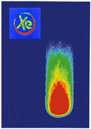
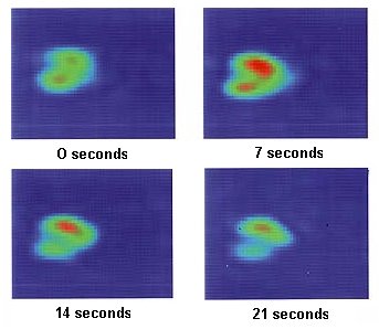
In recent experiments, Pines, Budinger and colleagues have used injections of xenon to deliver high concentrations of the gas directly into tissue. These MRI time-resolved images of a rat's upper hind leg show the effects after injection with laser-polarized xenon.
|
When spinning atomic nuclei align themselves with a magnetic field, their axes point in either an "up" or "down" direction. Because the signals from nuclei pointing in opposite directions cancel one another out, obtaining a useful MRI/ NMR signal depends upon an excess of nuclei being "polarized" in a single direction. Xenon nuclei show a small degree of natural polarization in their spin, but zapping them with a beam of polarized laser light creates a "hyperpolarized" effect in which most of the spins point in the same direction, a process based on findings from researchers at Princeton University.
Using a technique they call SPINOE (Spin-Polarization-Induced Nuclear Overhauser Effect), Pines and colleagues are able to transfer enhanced polarization from hyperpolarized xenon gas to other molecules in solution.
"Every area in a molecule or sample accessed by hyperpolarized xenon is going to light up to some extent with an enhanced nuclear magnetic resonance signal," Pines said when the SPINOE NMR technique was announced.
Xenon is particularly well-suited for medical studies. As an inert gas, its molecules can make physical contact with other types of molecules without affecting them chemically. Xenon is also readily absorbed in solutions, especially blood, but is repelled by water. It is expected that hyperpolarized xenon nuclei should huddle around water-free sites on proteins, increasing their influence on the spins of the protein's constituent nuclei which, in turn, would enhance the resulting MRI/NMR signal.
Pines, Budinger, and coworkers reported that introducing laser-polarized xenon directly into blood or tissue yielded high resolution time-resolved MRI/NMR signals. Their results point the way for using laser-polarized xenon gas as an alternative to radionuclides for imaging physiological phenomena in the blood system and in tissue specific to the heart, brain, and other organs.
| Lead scientist on this project
|
|
|
|
|
|
A major challenge was to find a means of efficiently delivering xenon to the blood and tissues while maintaining its enhanced polarization. The Berkeley Lab researchers accomplished this by pre-dissolving laser-polarized xenon in a "biologically compatible" solution. The polarized xenon gas was frozen at liquid nitrogen temperatures, sublimated, put into a solution, then shaken until it dissolved. As a result, loss of polarization during the delivery proved to be insignificant.
Until now, inhalation has been the primary method of delivering xenon to obtain detailed images of the lungs and brain. Recently, Pines and colleagues report that they have used injections of xenon to deliver high concentrations of the gas directly into tissue. This opens the possibility that physicians may someday choose inhalation or injection, depending on which organs they want to see.
- Lynn Yarris