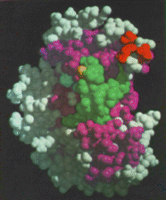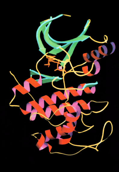
Scientists from LBL's Structural Biology Division, in collaboration with colleagues at UC Berkeley and UC San Francisco, have achieved an important step toward realizing this strategy -- by solving the three-dimensional structure of a key protein that triggers the last step of the events which culminate in cell division. This is the first time the structure of a protein involved directly in controlling cell division has been determined.
Cyclin-dependent kinase 2 (CDK2) is a member of a large family of protein kinases that initiates the principal transitions of the eukaryotic cell cycle, attaching phosphate groups onto their target enzymes. Various types of kinases are involved in the chain of reactions that follow cell stimulation by a growth factor. The critical step in the sequence of events is known as Start, the moment when the cell becomes committed to a new division cycle. It is at this stage when cell division can go awry, leading to cancer.
The three-dimensional structure of CDK2 was determined by Sung-Hou Kim's x-ray crystallography group at LBL -- an effort led by Hendrik L. De Bondt and Jarmila Jancarik. David Morgan led the San Francisco group in isolating and purifying the protein. The researchers collaborative effort was recently featured in an issue of the journal Nature .
X-ray crystallography merges art with science by growing a crystal from the protein of interest and then shooting it with an x-ray beam. The process generates distinct patterns which can be analyzed by computational methods to reveal a molecular structure. Computer graphics serve as a powerful tool to visualize the molecular surface and highlight possible targets for inhibitor design.
With the three-dimensional structure of CDK2 now determined, several research groups, including some biotechnology companies, are beginning to design potential drugs that bind and inactivate this protein.
This powerful tool, soon to be enhanced by beams of incomparable brightness generated by the Advanced Light Source, will provide promising opportunities for industrial linkages, furthering the investigation of structure-based drug design.