BERKELEY -- Some of the more shadowy secrets of biology may soon be
illuminated through the use of a new type of fluorescent probe developed by scientists
with the U.S. Department of Energy's Lawrence Berkeley National Laboratory and the
University of California at Berkeley.
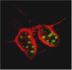 In this cross-section of mouse cells
labeled with two different sizes of semiconductor nanocrystals, nuclei show up as green
and actin fibers show up as red under the same illumination.
|
A joint LBNL-UCB research team led by Paul Alivisatos and Shimon Weiss has announced
the development of nanometer-sized crystals of semiconductors, such as cadmium selenide
and cadmium sulfide, that can be used as fluorescent probes for the study of biological
materials. These semiconductor nanocrystals offer a distinct advantage over conventional
dye-molecules in that they emit multiple colors of light, which means they can be used to
label and measure several biological markers simultaneously. The unique optical properties
of these semiconductor nanocrystals also hint at the possibility of observing changes that
take place in labeled biological systems, such as living cells, over a period of time.
This research was reported in the September 25, 1998 issue of the journal Science.
Alivisatos is a leader in the production by chemical means of semiconductor
nanocrystals, simple inorganic solids consisting of a hundred to a hundred thousand atoms.
He holds a joint appointment as a UCB professor of chemistry and a senior staff scientist
with Berkeley Lab's Materials Sciences Division (MSD). Weiss, a staff scientist with MSD,
is an authority on single molecule fluorescence and spectroscopy. He approached Alivisatos
with the proposal for this joint study.
Other members of the team who co-authored the paper in Science were Marcel Bruchez Jr.,
who also holds a joint LBNL-UCB appointment, and Mario Moronne and Peter Gin, with
Berkeley Lab's Life Sciences Division.
"Form follows function" is the golden rule in cell biology, which is why
microscopy has been the heart and soul of this research field and fluorescent-labeling one
of its most widely used tools. In fluorescent labeling, markers, usually antibodies that
attach themselves to specific proteins, are tagged with dye-molecules that fluoresce or
emit a specific color of light when stimulated by laser light, usually from a confocal
microscope.
"Sometimes, in order to fully characterize a sample, a population of cells, for
example, you need to look at combinations of markers," says Alivisatos. Such
measurements require multiple-color light emissions which are difficult to obtain with
conventional dye molecules.
"Ideal probes for multi-color experiments should emit at spectrally resolvable
energies, should have a narrow, symmetric emission spectrum, and the whole family should
be excitable at a single wavelength," the authors of the Science
paper wrote.
Semiconductor nanocrystals met these demands in a "dual emission from single
excitation" labeling experiment on mouse tissue cells called 3T3 fibroblasts. A core
nanocrystal of cadmium selenide was enclosed within a shell of cadmium sulfide to boost
the amount of fluorescence and reduce photochemical degradation. This core-shell combo was
then enclosed within a shell of silica for water solubility and biocompatibility.
With earlier work by Alivisatos having shown that the color of light emitted by a
semiconductor nanocrystal depends upon its size, the mouse cells were labeled with two
different sizes of core-shell nanocrystals. It was also known that modifying the surface
of the silica shell can be used to selectively control its attachment to components within
a cell. In this case, the smaller nanocrystals (two nanometers), which fluoresced green,
were modified to penetrate the nucleus of each cell, and the larger nanocrystals (four
nanometers), which emitted red light, were modified so that they would attach themselves
to actin filaments along the outer cell membrane.
Using wide-field microscopy, the green and red labels were clearly visible to the naked
eye and could be photographed in true color with an ordinary camera. Confocal microscopy
images showed that cell nuclei had been penetrated with the green probes and the actin
fibers had been stained red. After repeated scans, the nanocrystal labels showed far less
photobleaching than would occurred in the control sample labeled with conventional dye
molecules.
"The development of semiconductor nanocrystals for biological labeling gives
biologists an entire new class of fluorescent probes for which no small organic molecule
equivalent exists," the authors of the Science paper wrote.
"These nanocrystal probes can be complementary and in some cases may be superior to
existing fluorophores."
The authors also assert that, compared with conventional fluorophores, semiconductor
nanocrystals have a "narrow, tunable, symmetric emission spectrum, and are
photochemically stable." These features, along with a relatively long fluorescence
lifetime (hundreds of nanoseconds) indicate that, in addition to serving as direct probes,
semiconductor nanocrystals could also be used as "sensitizers" for traditional
dye-molecule probes, meaning they transfer their excitation energy to the dye-molecule.
An earlier research team led by Weiss demonstrated that this energy sensitizing
phenomenon, known as fluorescence resonance energy transfer (FRET), when it takes place
between a single donor and a single receptor, could allow for the labeling and observation
of dynamic events such as conformational changes in a protein.
"The use of semiconductor nanocrystals should allow us to do unique FRET
experiments," says Weiss. "For example, labeled molecules could be made to emit
different colors at different times of an event."
Weiss and Alivisatos and their co-authors also believe that semiconductor nanocrystals
could be applied to x-ray- and electron-based imaging techniques, and could serve as
tunable infrared dyes for detecting fluorescence in, among other things, blood samples.
Berkeley Lab is a U.S. Department of Energy national laboratory located in Berkeley,
California. It conducts unclassified scientific research and is managed by the University
of California.
