| February 17, 2004 | science beat | | lab a-z index | lab home |
|
|||
| Glass Beads Reveal Molecular Interactions | |||||
| Contact: Dan Krotz, dakrotz@lbl.gov | |||||
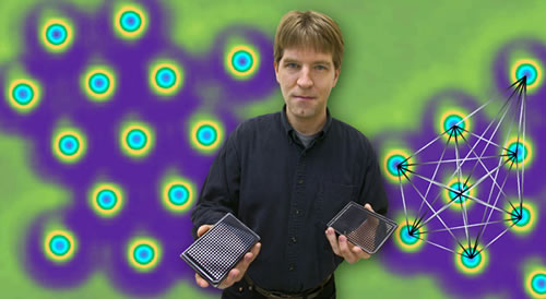
| Berkeley Lab and UC Berkeley researchers have developed a fast, cheap, and highly sensitive way to detect molecular interactions without using sophisticated equipment. Their technique, which uses thousands of microscopic glass beads coated with a substance that mimics a cell membrane, opens the door for the high throughput evaluation of an ever-growing family of pharmaceuticals that fight diseases by targeting membrane-bound receptors.
As described in a recent issue of Nature, the technique takes advantage of the unique properties of a colloid, a substance consisting of tiny particles suspended in a liquid. Given the right conditions, a slight change in energy can trigger a colloid to undergo a phase transition, in which short-lived clumps of particles disperse like the opening shot of a pool game. In this case, the tiny particles are silica beads, and when proteins bind to receptors embedded in their membrane-like outer layer, the beads scatter—a phenomenon that can be easily seen with a microscope. "We position the colloid to teeter on the edge of a phase transition, so the slightest change, like the binding of proteins to receptors, causes the beads to spread out," says Jay Groves of the Physical Bioscience Division and professor of chemistry at UC Berkeley, who conducted the research with fellow UC Berkeley chemists Michael Baksh and Michal Jaros. In this way, something as minute as a molecular interaction sparks a major change in how the entire population of ever-moving beads aggregates. This advantage, called signal amplification, means protein-receptor interactions can be detected with a low-magnification microscope, and this means drug candidates that target protein receptors can be evaluated with a fully automated process. "The colloid system can be implemented into existing high throughput screening protocols," says Groves. Other ways of determining how proteins latch onto cell-membrane receptors involve labeling molecules with fluorescent markers, which is very costly and time-consuming, or using sophisticated equipment like a surface plasmon resonance spectrometer, which is not very sensitive. Because of their drawbacks, the techniques loom as a bottleneck in the evaluation of thousands of molecules that could potentially inhibit a receptor's ability to bind with disease-causing proteins. For example, approximately 60 percent of today's prescription drugs target a class of signaling receptor called a 7-transmembrane helix protein. Although these drugs represent a huge market, averaging $100 billion annually, researches have so far only determined how a small minority of the myriad types of 7-transmembrane helix proteins bind to receptor-specific molecules, also called ligands. As Groves explains, there is tremendous interest in finding ligands that bind with the remaining majority of poorly understood transmembrane proteins, or so-called orphan proteins. "This would significantly advance development in this well-established goldmine of drug targets," says Groves. What's needed is a high-throughput way of exploring these ligand-protein interactions, both to identify the ligands and to screen for molecules that compete with them. Groves envisions a process in which hundreds of colloid suspensions are created, each composed of glass beads coated with membranes containing different types of 7-transmembrane helix proteins. Next, a robot dispenses these colloid clusters into tiny wells, and potential ligands or ligand competitors are then added to each well. To determine if an interaction occurs, the colloids are scanned by an automated microscope designed to image thousands of samples in rapid succession. If a colloid's beads scatter—something computer-assisted image analysis can detect—then the ligand or ligand competitor has latched onto the transmembrane protein. "The entire process wouldn't require sophisticated instruments," says Groves. "High-throughput systems, such as those used by pharmaceutical companies, could run our assay at a rate of 100,000 measurements per day." In addition to expediting drug development, the technique could be used to diagnose the presence of membrane-targeting toxins such as anthrax, botulism, cholera and tetanus. And because the majority of biochemical processes occur on cell membranes, it can also be applied to fundamental cell biology research. Groves' colloid technique isn't his first foray into simplifying the study of cell membranes. His MembraneChip™, a device developed a few years ago, detects protein-receptor interactions by attaching a cell membrane to a silicon electronic chip. "I saw in these colloids a faster variation to the MembraneChip," says Groves. "And instead of powering the device with electricity, colloids use ambient thermal energy. It's a power-free, label-free, and instrument-free system." Additional information
|
|||||
| Top | |||||