| science@berkeley lab | | lab a-z index | lab home |
|
|||
| The Language of Magnetic Resonance Part 2: Quieter and More Versatile Laser-detected Magnetic Resonance Imaging |
|||||||||||||||||||||||||||||||||||
| Contact: Lynn Yarris, lcyarris@lbl.gov | |||||||||||||||||||||||||||||||||||
Magnetic resonance imaging, MRI, is synonymous with huge doughnut-shaped machines, loud scary noises, and signs that warn of magnetic fields so powerful patients must remove watches, jewelry, and all other metallic belongings before a scan. A far less intimidating alternative is in the offing with recent advances made by chemist Alex Pines — one of the world's foremost authorities on MRI and its sibling, nuclear magnetic resonance (NMR) — and his celebrated "Pinenuts" research group.
Called Laser-detected MRI (LMRI), this latest development from Pines and his group is based on a combination of remote NMR/MRI signal detection and optical atomic magnetometry. LMRI dispenses with the need for powerful magnets — in fact it can even use Earth's weak magnetic field — as well as the complex cooling schemes that are also typical of conventional NMR/MRI systems. So promising is the new LMRI technology that it won an R&D 100 Award for 2007. Established by R&D Magazine to recognize "the most significant proven technological advances of the year," R&D 100 Awards have been dubbed the Oscars of technology. "I believe we were the only group to win an R&D 100 Award this year for a project that is fundamental scientific research," says Pines, who holds joint appointments with Berkeley Lab's Materials Sciences Division and with UC Berkeley, where he is the Glenn T. Seaborg Professor of Chemistry. "Our LMRI technique is basically simple and does not involve any expensive components, yet it provides a viable MRI signal [and] enhanced sensitivity and time resolution, for situations where traditional MRI is not practical or in some cases not even an option." LMRI offers a broad range of potential applications and can be used on samples as vast as petroleum reservoirs or as tiny as cellular tissue samples. Of all the potential applications of LMRI, perhaps the most important is its possible use in functional magnetic resonance imaging (fMRI) systems. Since its invention in the early 1990s, fMRI has become one of the primary diagnostic tools for identifying and characterizing neurological damage and disorders. It is noninvasive, quick, and does not involve ionizing radiation that can damage cells. Unlike the static images captured by conventional MRI, fMRI brain scans produce a series of images that can reveal changes in cerebral blood flow during neural events. The magnetic properties of blood change when it is oxygenated, and the degree of oxygenation, along with blood flow (collectively known as hemodynamics), is closely linked to specific neural activities. However, because of the powerful magnetic field required for conventional fMRI scans, the technology cannot be used on patients with shrapnel, bullet fragments, or other metal objects lodged inside the brain. "We've shown that with LMRI, we can detect the flow of blood or other fluids that contain magnetic particles with no external magnetic field — that is, we can run fMRI scans using the magnetism only from Earth's field," says David Michalak, one of the members of Pines's group now working on the project. "So far we've only tested this on gas flows through a polystyrene brain. But down the road we can envision a system in which you could prepolarize a fluid and introduce it into the brain, let it flow through a region of interest and into the internal jugular, where you could collect an MRI signal. This should be possible within the next five years."
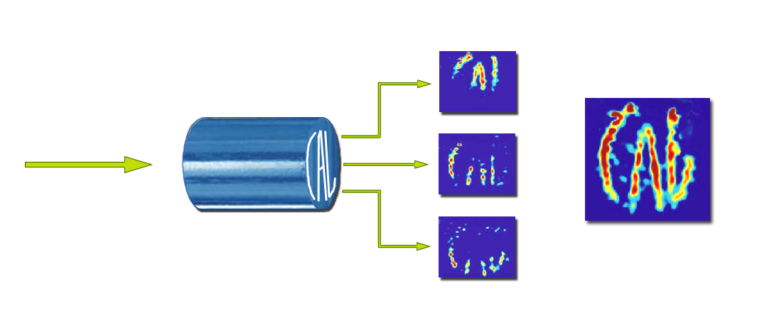
Another extremely important application of LMRI is in the burgeoning field of microfluidic devices. These "laboratories on a chip" feature an array of micrometer-sized channels and ports etched into a chip for moving and analyzing nanoliter-sized samples of fluids, which can provide a wealth of information for biomedical and analytical chemistry studies. Because of their incredibly small sample sizes — samples that are thousands of times smaller in volume than a typical droplet — microfluidic devices are highly prized for providing rapid analysis at relatively low costs. Today's microfluidic chips rely on marker particles, however, which are added to the flow and either fluoresce under optical illumination or can be viewed with a microscope. Such markers, which can perturb fluid flow, are not needed with LMRI. A Parting of the WaysThe foundation for LMRI began with the separation of the two basic steps to NMR/MRI technology — signal encoding and signal detection. In a conventional NMR/MRI setup, these two processes take place within a single device. Two years ago, Pines and his group devised a format in which these processes are physically separated and carried out independently. This approach, called remote detection, opened the door to LMRI and its application to microfluidic chips. "Not only did our methodology bypass the long-standing problem of optimizing signal encoding and detection, it also added an important new dimension to the study of fluid flow dynamics, which was time-of-flight measurements," says Pines. "In addition to time-of-flight measurements, remote NMR/MRI detection overcame traditional sensitivity limitations and enabled spatially resolved imaging." Christian Hilty, another member of Pines's research group, was the principal author of a paper in the Proceedings of the National Academy of Sciences that first announced the remote NMR/MRI detection technique. "It offers the unique advantage of being non-invasive," Hilty says. "We can use remote NMR/MRI detection to measure microfluidic flow without the introduction of foreign tracer particles, which is an important advantage for the design and operation of microfluidic devices."
NMR/MRI is based on spin, a property of atomic nuclei in which they act as bar magnets with a north and south pole. When exposed to a strong external magnetic field, the nuclei attempt to align their axes along the lines of magnetic force. Since the alignment is not exact, the result is a wobbling rotation, or precession, that's unique to each type of atom. If while exposed to the magnetic field the precessing nuclei are also subjected to radiofrequency (rf) pulses, they will absorb and re-emit energy at characteristic frequencies. This is the signal-encoding phase of NMR/MRI. In the detection phase, the frequencies of the encoded signals are either measured to obtain a spectrum, as in NMR, or used in MRI to obtain a spatially encoded signal that can then be translated into images. Obtaining a useful NMR/MRI signal from a sample depends upon polarizing the spins of its precessing nuclei so that an excess of spins point in one direction, either "up" or "down." Conventional NMR/MRI technology uses an exceptionally strong external magnetic field to produce a detectable signal. The stronger the magnetic field, the stronger the signal, which in the past has meant a large, expensive, cryogenic, high-field magnet.
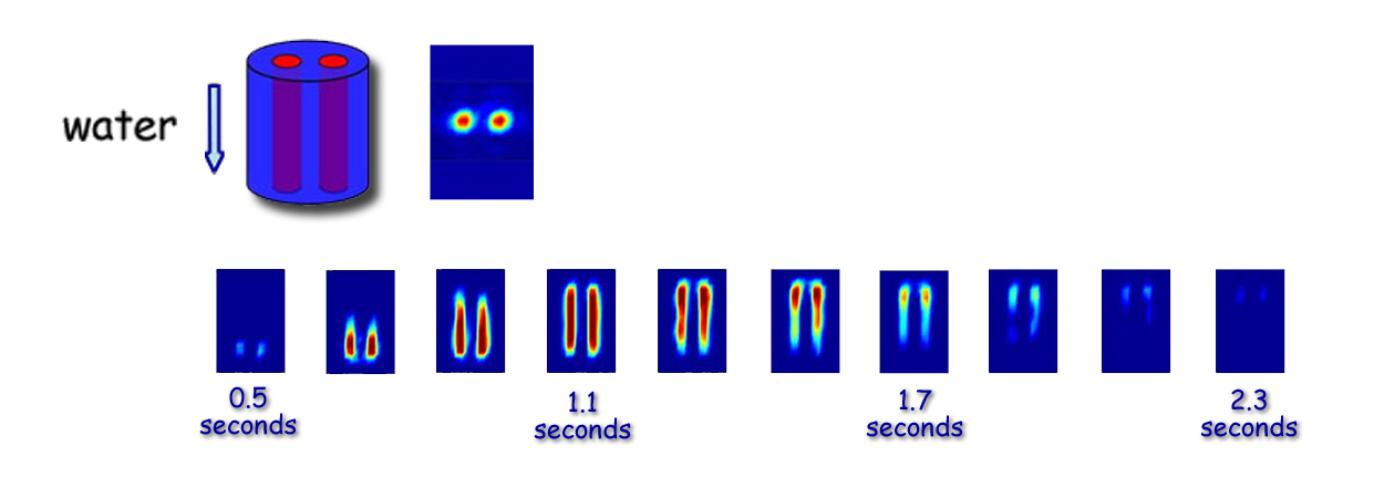
In the initial development of their remote NMR/MRI technique, Pines and his research group eliminated the need for an expensive cryogenic high-field magnet by strengthening their NMR/MRI signals with a xenon gas additive whose atomic nuclei were hyperpolarized by laser light. In transferring the hyperpolarization to hydrogen and other atomic nuclei, the xenon nuclei can boost the NMR/MRI signal of a sample by at least four orders of magnitude, even in Earth's magnetic field. Xenon being chemically inert, the addition of xenon nuclei does not otherwise affect a sample's constituents. Since it takes several minutes for hyperpolarized xenon nuclei to lose their spin polarization, samples encoded with NMR/MRI information in this manner can be transported to a separate site for detection. Once they were able to carry out the encoding and detection phases of NMR/MRI at separate sites, Pines and his group could customize each phase to obtain optimal results. This opened the door to making NMR/MRI technology compact, portable, relatively cheap — and a whole lot quieter. Optical Atomic MagnetometryEncoding a sample with an NMR/MRI signal using only the Earth's magnetic field — even when the sample is prepolarized — results in a signal that is weaker than the signal from conventional high magnetic field encoding. Consequently, sensitivity in the signal detection phase of the process must be greater. One of the means by which Pines and his group are boosting detection sensitivity is through the use of a unique variation of optical atomic magnetometry. Magnetometers are devices used to measure the strength and direction of a magnetic field. The optical atomic magnetometer used by Pines and his research group was developed by Dmitry Budker, a physicist who holds a joint appointment with Berkeley Lab's Nuclear Science Division and UC Berkeley's Physics Department. Budker's version of a magnetometer is based on a phenomenon called nonlinear magneto-optical rotation, which endows his device with high sensitivity to low-field magnetic signals at room temperature. "The fact that we can detect a viable signal without the use of superconducting magnets or cryogenics significantly reduces the cost and maintenance of the apparatus, and opens the technology up to a broader range of applications," says Shoujun Xu, a member of Pines's research group who conducted the first MRI measurements with Budker's magnetometer. "Furthermore, our technique has simple electronics that can be easily integrated into detector arrays." With the optical atomic magnetometer, a sample of alkali atoms featuring a single unpaired electron is vaporized in a glass cell. This endows the alkali atoms with a magnetic moment three orders of magnitude stronger than that of precessing nuclei. A beam of laser light is then used first to pump the atoms so that their spins are polarized, then to probe the polarized atoms for an MRI signal. According to Budker, instead of the multimillion-dollar costs of a conventional MRI system, LMRI technology costs only a few thousand dollars to implement. Instead of weighing several tons, the equipment weighs less than 100 pounds. "Our system is fundamentally simple and does not involve any single expensive component," Budker says. "We anticipate that the whole apparatus will become quite compact, and deployable as a battery-powered, portable device." In the LMRI system developed by Pines and his group, a fluid to be imaged is passed through two small cells for signal encoding, then transported to a U-shaped detection area for interrogation by a pair of Budker's magnetometers. The magnetometers are oriented so that they detect the MRI signal with opposite signs. This configuration dramatically improves the signal-to-noise ratio, enabling the researchers to detect MRI signals from microliters of sample in a split second, without the presence of a strong magnet. "Being able to use LMRI in Earth's magnetic field offers a number of key advantages over traditional high-field MRI," says Pines. "The entire signal-encoding setup is portable; the encoding volume can be as big as any object to be imaged" — something not possible when superconducting magnets are required — "electrical power and rf requirements are modest, and Earth's magnetic field is always available." In addition to its potential use in fMRI brain scans and microfluidic chips, LMRI has been demonstrated to have potential for monitoring distributions of magnetically labeled DNA and protein molecules or antibodies in the immune system. Pines and his group have also shown that LMRI can detect certain phenomena involving atomic nuclei that are characteristic of the formation or cleavage of chemical bonds. "The ability to detect even single particles in large amounts of sample should also be valuable for security applications, as LMRI could be used to screen for magnetically labeled viruses in dilute environments. Or it could be used as a means of inline quality control for industrial processes involving magnetic products or impurities," says Pines. "LMRI should also make it possible to study biochemical events that are associated with the aggregation of magnetic particles."
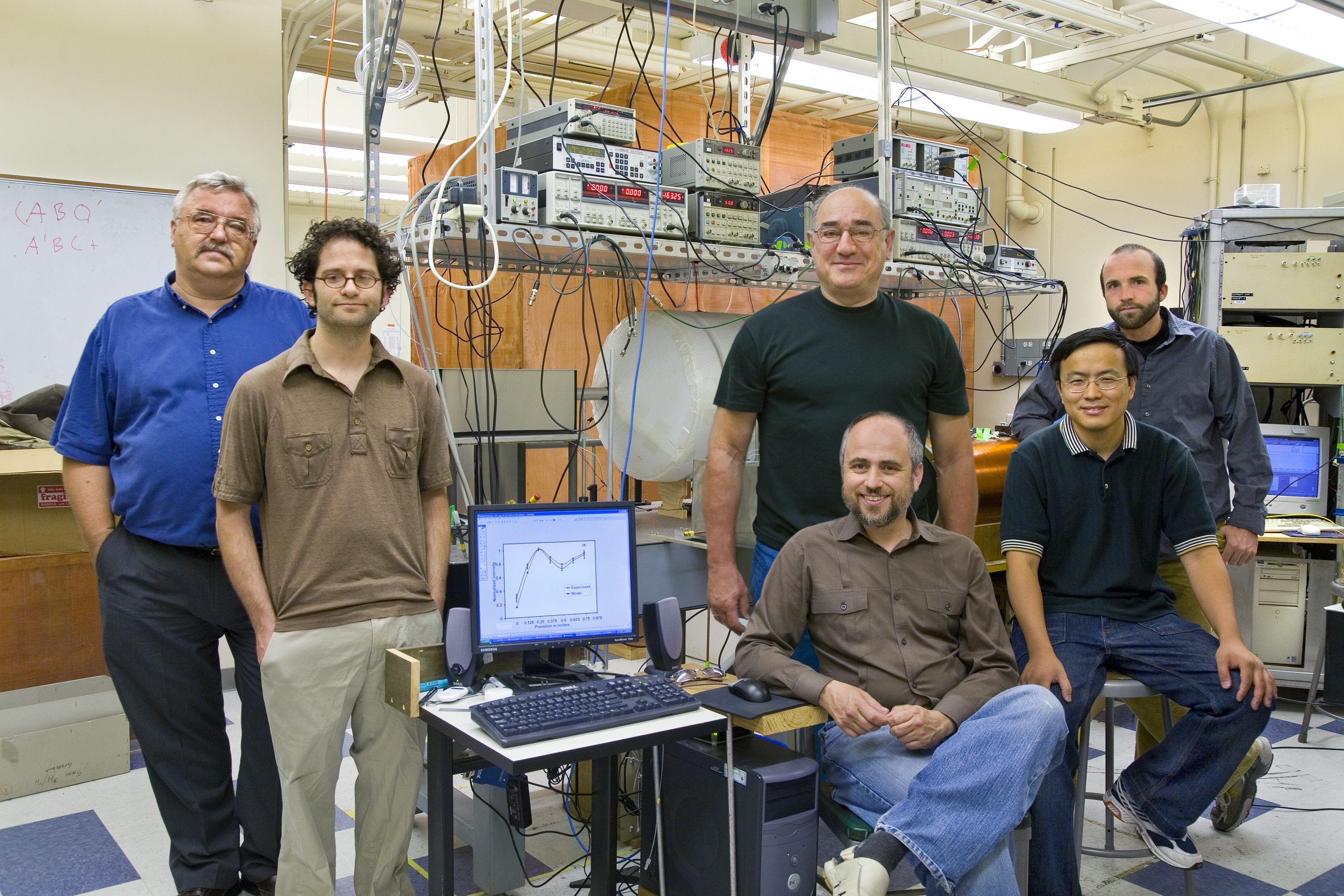
In addition to Pines, Budker, and Xu, others who shared in the 2007 R&D 100 Award for LMRI technology were Marcus Donaldson, Simon Rochester, and Valeriy Yashchuk. Additional information
|
|||||||||||||||||||||||||||||||||||
| Top | |||||||||||||||||||||||||||||||||||