| May 30, 2006 | science@berkeley lab | | lab a-z index | lab home |
|
|||
| Paving the Way for Early Detection of Cancer | ||||||||||||||||||||
| Contact: Dan Krotz, dakrotz@lbl.gov | ||||||||||||||||||||
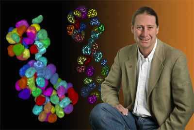
A diagnostic test that exposes the first inkling of cancer in a cell's nucleus could someday become a reality, thanks to a team of Berkeley Lab and Purdue University scientists who have developed a way to automatically map the three-dimensional distribution of proteins within the nuclei of human mammary cells.
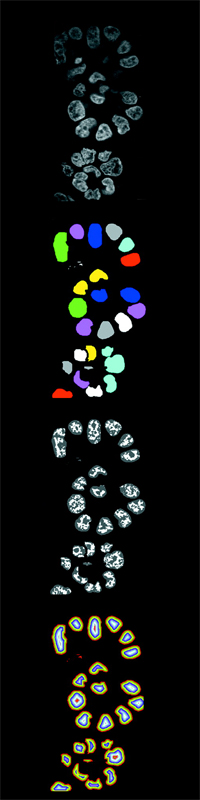
The technique, which couples state-of-the-art fluorescent imaging with computer-driven image analysis, could enable doctors to detect aggressive cancers in their earliest stages — giving physicians more time to fight the disease. It could also allow doctors to better characterize a patient's cancer and match him or her with the most effective therapy. "It won't replace today's tests, but it will give pathologists an important additional view of tissue," says David Knowles, a scientist in Berkeley Lab's Life Sciences Division who pioneered the system with Sophie Lelièvre. Lelièvre, a former postdoc in the laboratory of Berkeley Lab Distinguished Scientist Mina Bissell, is now an assistant professor of Basic Medical Sciences at Purdue University. Their technique hinges on the fact that inside a cell's nucleus, proteins arrange themselves in a different pattern depending on whether the cell is malignant, hasn't developed a specific function, or is perfectly healthy and mature. In the case of malignant cells, this telltale shift in protein distribution is believed to occur before the cells proliferate. It's as if cells destined to become cancerous tip their hand to anyone clever enough to read it, which is where Knowles and Lelièvre come in. First, they stained breast cell samples with a fluorescent substance that targets a protein, called NuMA, which is found in the nuclei of human mammary cells. When functioning properly, the protein helps regulate various aspects of the cells' health. But when it malfunctions, the protein has been linked to leukemia and breast cancer. Under confocal microscopy, the tissue samples appear as if they are painted with a large swath of luminescence, indicating the presence of thousands of nuclei and their NuMA proteins. Next, to pinpoint the spatial distribution of the proteins within each nucleus, they applied image analysis software developed by Knowles. The software automatically delineates the boundaries of each nucleus in a tissue sample, and then homes in on the brightest spots of luminescence within each nucleus — revealing the precise location of each protein. A final step allows the scientists to map the distribution of the proteins by digitally unraveling the nuclei, starting from the outer edges and working toward their centers. "We peel away the layers, like an onion, and quantify the proteins in each layer," says Knowles.
Their analyses, which were conducted on tissue samples developed in the lab, reveal that the distribution of NuMA is very different in malignant cells compared to both differentiated cells (cells that have become specialized for a particular function) and nondifferentiated cells. "In malignant cells, we find that the organization of the proteins is haphazard and completely disrupted," says Knowles. "Whereas with the nonmalignant cells, there is a real organization pattern, which changes as the cells differentiate." Knowles and Lelièvre can even see the difference between malignant cells and nondifferentiated, nonmalignant cells that proliferate, which means they can identify cancer-related characteristics that are very difficult or impossible to measure with usual cell biology techniques. Although their technique is still in the early developmental stage, it could also contribute to the current thrust in breast cancer treatment, in which scientists are working to understand the different shades of cancer and target each shade with specially tailored therapeutics. This method requires a degree of characterization that today's techniques can't always deliver. For example, pathologists currently examine potentially cancerous tissue samples using techniques such as Hematoxylin and Eosin staining. Although this workhorse test will likely be used for decades to come, it is a broad-brush tool that mainly captures changes in the shape and spatial distribution of cells and nuclei. If changes to smaller-scale phenomena are captured, such as the distribution of individual proteins, then pathologists will able to better characterize cancer — and attack it with the best treatment. "If we could play a role in this, we would be ecstatic," says Knowles. "A pathologist could conduct our analysis automatically over a large population of cells and map the results back onto the tissue. It would represent an entirely new platform on which they can base their decision." So far, Knowles and Lelièvre have only used the technique to explore how changes to one nuclear protein, NuMA, impact the malignancy of a cell. In the future, they'd like to broaden their inquiry by mapping a range of proteins and determining if changes to their spatial distribution are also a hallmark of cancer. Ultimately, their technique may join the many tools physicians use to diagnose and characterize cancer, such as genetic tests, medical imaging, and Hematoxylin and Eosin staining. "The end goal is to combine these methods," says Knowles. "Our technique could augment a pathologist's report and shed a new light on how cancer is characterized." Their research is published in a recent issue of the Proceedings of the National Academy of Sciences. In addition to Knowles and Lelièvre, Carol Bator-Kelly of Purdue University and Berkeley Lab's Damir Sudar and Mina Bissell contributed to the research. Additional information
|
||||||||||||||||||||
| Top | ||||||||||||||||||||