| September 23, 2005 | science@berkeley lab | | lab a-z index | lab home |
|
|||
Breast Cancer Research at Berkeley Lab
|
|||||||||||||||||||||||||
| Contact: Lynn Yarris, lcyarris@lbl.gov | |||||||||||||||||||||||||
| The final installment in a series on research at Berkeley Lab that aims to eradicate the scourge of breast cancer. Tumors originate in cells that are actively dividing, a fact well-established by cancer researchers. The thought is that such cells are more likely to acquire genetic mutations that can lead to cancer. Even though cell division activity is highest as we mature from childhood into adulthood, however, cancer is a disease primarily associated with older adults.
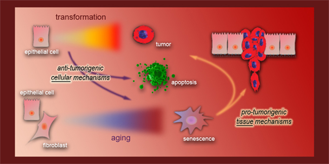
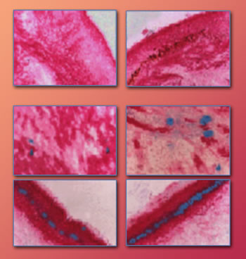
One possible answer for this conundrum may be found in cellular senescence, the process by which cells stop dividing in response to stress or damage. That's the hypothesis being pursued by Judith Campisi, a cell biologist in Berkeley Lab's Life Sciences Division, a cofounder of the Center for Research and Education on Aging (CREA), and a leading authority on cell senescence and the effects of aging. In a presentation prepared for the 2005 Era of Hope conference on breast cancer, held last June in Philadelphia (see Part 1 of this series), Campisi offered evidence that cellular senescence is a double-edged sword: it helps protect children and young adults from developing cancer but becomes a contributing factor to the development of cancer later in life. "We have proposed that cellular senescence is an example of evolutionary antagonistic pleiotropy," Campisi said. "While senescence response benefits young organisms by suppressing the development of cancer, the persistence of dysfunctional senescent cells can disrupt normal tissue structure and function and thus contribute to aging and age-related pathology, including cancer." The Era of Hope conference posed the question Where are we in the conquest of breast cancer? For all of the advances made in diagnostics and treatments, breast cancer remains one of the deadliest forms of the disease. In the United States, breast cancer strikes one out of every eight women and kills one out of every 33 women it strikes. All forms of cancer arise from cell division run amok. Nature has evolved two ways of shutting down the cell-division process and reining in this uncontrolled growth: senescence, in which cells remain metabolically active but are no longer are capable of dividing, and apoptosis, or cell suicide. The role of apoptosis as a tumor suppressor has been well characterized through in vivo studies, but the role of senescence in living organisms has not been so clearly defined. Although cellular senescence was first reported in cultures some 40 years ago, it was not until 1995, when Campisi and a group of collaborators announced the development of a fast and simple in vivo assay, that scientists could even confirm that senescence occurs in living organisms. Campisi's assay was based on the discovery that senescent cells express an unusual form of an enzyme called beta-galactosidase, which is generally absent in presenescent cells. The assay she and her collaborators developed employed a stain that turns blue in the presence of beta-galactosidase.
Based on studies of senescent cells in mice, Campisi hypothesized that over time an organism acquires enough senescent cells to create a procarcinogenic microenvironment. In such an environment, conditions would be conducive to transforming benign lesions into malignant tumors. "There is increasing evidence that genetic mutations alone are insufficient for cancer development," she said in her presentation at the Era of Hope conference. "Rather, malignant tumors also require a permissive tissue environment in which to develop and progress. We propose that the accumulation of senescent cells, which can disrupt normal tissue structure and function, is an independent synergistic process that contributes to cancer progression." This hypothesis, Campisi said, could help explain why tumor incidence increases exponentially with age. In addition to beta-galactosidase, senescent cells secrete other enzymes that degrade nearby tissue. The effects are readily apparent when you compare the smooth skin of a child to the wrinkled skin of a senior citizen. "Age-related decrements in tissue may, at least in part, derive from an accumulation of senescent cells which cannot proliferate, resist apoptotic death, and have an altered phenotype," said Campisi. Working with human and mouse mammary epithelial cells and the stromal cells that support them, grown in two- and three-dimensional cell culture systems, Campisi and her research group have shown that senescent stromal cells can stimulate the proliferation of normal and precancerous mammary epithelial cells. In addition, senescent cells can disrupt the functional and morphological differentiation of mammary epithelial cells, specifically their ability to produce milk proteins and form ductal branches. "We've begun to characterize the phenotype of senescent breast epithelial cells, about which little is known, and explore the influence of senescent mammary epithelial cells on nonsenescent normal or precancerous neighboring cells," Campisi said. "We have found that the replicatively senescent state of human mammary epithelial cells, like human stromal cells, is accompanied by very little apoptosis, which can, in part, explain their stability and persistence."
In their tests, Campisi and her colleagues showed that when they turned off the tumor-suppressing protein known as p53, senescent mammary epithelial cells could be induced to resume the cell division process. However, when this was done, the incidence of apoptosis increased. The researchers also showed that senescent mammary epithelial cells, like senescent stromal cells, can interfere with the ability of nonsenescent breast cells to undergo morphological differentiation. They are now studying the enzymes secreted by senescent human breast cells to identify candidate proteins that may be responsible for the effects on their nonsenescent neighbors. The link between the prolonged presence of senescent cells and the development of malignant tumors could provide new ways to treat breast cancer, if therapies can be developed that either eliminate senescent cells in older women or prevent senescent cells from secreting enzymes that damage their neighbors. Campisi says, "In theory, if you can get senescent cells to die, you can retard cancer. You'd still have mutations, but you wouldn't have the all-important synergy between mutations and cellular senescence." Additional information
|
|||||||||||||||||||||||||
| Top | |||||||||||||||||||||||||