| September 23, 2005 | science@berkeley lab | | lab a-z index | lab home |
|
|||
Why Older People Suffer More Bone Fractures
|
|||||||||||||||
| Contact: Dan Krotz, dakrotz@lbl.gov | |||||||||||||||
| It's no mystery that elderly people are more prone to bone fractures than younger people. What is a mystery is precisely why. Now, Berkeley Lab scientists have gained a better understanding of this problem using state-of-the-art imaging, testing techniques and detailed mechanical analysis. They found that as people grow older, their bones lose their ability to resist the formation and growth of cracks that lead to breaks. This insight into the mechanics of bone fractures could help scientists develop new ways to fight the effects of aging, injury and disease.
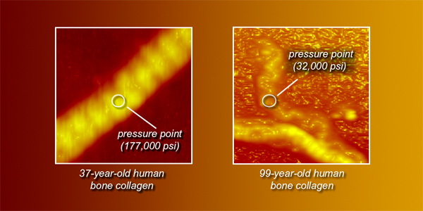
"It is my hope that by understanding in detail the mechanisms by which healthy bone resists fracture, new therapeutic measures can be developed that will help preserve bone strength as people age," says Robert Ritchie, a scientist with Berkeley Lab's Materials Sciences Division and chairman of UC Berkeley's Department of Materials Science and Engineering. It's widely known that people are more susceptible to bone fractures as they age, sometimes with dire consequences. Half of women over age 50 and 25 percent of men over 50 will have an age-related bone fracture sometime in their lifetime. Doctors have traditionally pinned this on the fact that bone mineral density decreases with age. But several recent studies have failed to find a direct link between changes in bone mineral density and risk of fracture, indicating that other factors may be involved. Perhaps changes in bone toughness — or how well a bone's structure resists fracture — is also a culprit. In other words, bone quality may play as much a role in fractures as bone quantity. "Obviously it's not good to lose bone mass, but we think that it's only a part of the story. We need to really understand what happens to the structure of bone and how this affects its resistance to fracture," says Ritchie. To do this, Ritchie and fellow Materials Sciences Division scientists Joel Ager, postdoctoral fellows Ravi Nalla and Jamie Kruzic, and John Kinney of Lawrence Livermore National Laboratory turned to the same resources they've used to conduct pioneering research on advanced materials, composites, and ceramics. These include mechanical testing techniques that examine how cracks behave in materials, and two Berkeley Lab user facilities that yield extremely high resolution images: the Advanced Light Source, where the nation's premier source of synchrotron x-rays enables tomographic studies of materials, and the National Center for Electron Microscopy, where scientists conduct transmission electron microscopy characterizations of materials. "We've been able to bring to bear Berkeley Lab's absolutely state-of-the-art materials characterization techniques to help us better understand bone fracture," says Ritchie. The team used these tools to examine human cortical bone from cadavers that ranged in age from 34 to 99 years. Because bone is structurally unique at different scales, they examined the bone at the macroscale down to the nanoscale, where mineralized collagen fibers measure 50 to 70 nanometers in diameter ( a nanometer is one billionth of a meter). They found that the resistance of bone to the initiation of cracks falls by 40 percent over the age range examined. Most strikingly, among the very elderly, cortical bone completely loses the ability to resist the growth of an existing crack.
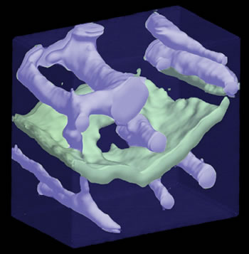
The reason for this sharp drop-off in fracture resistance plays out on several size scales. Three-dimensional x-ray computed tomography images obtained at the Advanced Light Source and the Stanford Linear Accelerator Center reveal that the bones of younger people have large unbroken regions of bone between cracks. These unbroken "bridges," which span tens of micrometers ( a micron is a millionth of a meter ), act like steel rods in reinforced concrete. In older people, however, the amount of these unbroken crack bridges is greatly reduced, meaning that it's easier for one tiny crack to connect to another — the makings of a larger fracture. "We see cracks opening up ahead of other cracks, and the regions in between act as bridges," says Ritchie. At the nanoscale, ultraviolet Raman spectroscopy studies reveal changes in the cross-linking underlying the collagen fibrils. "With age, changes in the cross-linking of collagen fibrils stiffens the bone structure — much like fusing together a plate full of spaghetti," says Ritchie. "This can make the bone more brittle." Although a hierarchy of structures at various size scales contributes to bone failure, Ritchie and his coworkers found that microcracks appear to initiate at the boundaries of osteons, which are concentric rings of collagen fibrils that surround blood vessels in bone. These osteons measure on the order of ten to several hundred microns across. "This is the dominant dimension for fracture in bone and the scale of the microstructure that we think principally controls the fracture processes," says Ritchie. Ritchie believes this mechanistic understanding of bone failure could help inform the treatment of bone injuries and diseases such as osteoporosis. Currently, bone aging is treated in terms of a loss of bone quantity, but his team's research reveals that other factors also play a role. "Perhaps it's time for new therapeutic treatments," Ritchie says. "At one end there is a risk of fracture, and at the other end there is disease, aging, and injury. We're learning that structural changes link these two sides." This research is detailed in a number of recent publications, including the December 2004 issue of the journal Bone under the title "Effects of aging on the toughness of human cortical bone: Evaluation by R-curves." Additional information
|
|||||||||||||||
| Top | |||||||||||||||