| June 29, 2005 | science@berkeley lab | | lab a-z index | lab home |
|
|||
| Breast Cancer Research at Berkeley Lab
Part 1: An Era of Hope for Breast Cancer Patients |
|||||||||||||||||||||||||||||||||||||||||
| Contact: Lynn Yarris, lcyarris@lbl.gov | |||||||||||||||||||||||||||||||||||||||||
| Beginning a series on research at Berkeley Lab that aims to eradicate the scourge of breast cancer.
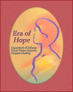
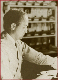
In June 2005, one of this country's premier conferences on breast cancer research took place in Philadelphia. Some 2,000 scientists, physicians, breast-cancer survivors, policy-makers, and advocates came together in the City of Brotherly Love to pool their knowledge and ideas about the ongoing search for improved diagnostics and treatments for the second leading cause of cancer death among women of all ages. Many readers might be surprised to learn that this meeting, called the 2005 Era of Hope, was sponsored by the U.S. Department of Defense. Through its Breast Cancer Research Program, DOD is the second largest funding agency for breast cancer research in the world. Readers might also be surprised to learn that the Department of Energy's Lawrence Berkeley National Laboratory had a strong scientific presence at the conference, with nearly two dozen researchers participating through either talks or posters. Berkeley Lab has been one of the pioneering institutions in cancer research, dating back to 1937 when John Lawrence, younger brother of the Lab's founder, Ernest Lawrence, used radio-isotopes produced at one of the Lab's cyclotrons to successfully treat their mother for cancer of the uterus. John Lawrence, who would become known as the "father of nuclear medicine," made the determination that neutrons, which have a destructive effect on living tissue, could be used to destroy tumor cells. In 1939 he began experimental treatments of several forms of cancer using neutron beams produced in particle accelerators. Enough progress was made over the next two decades that in 1954 he was able to launch the first heavy-ion stereotactic radiosurgery program. This program began with the use of proton beams produced at Berkeley Lab's famed 184-Inch Cyclotron to treat patients with breast cancer.
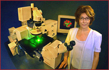
Berkeley Lab researchers continued to develop heavy-ion stereotactic radiosurgery as a treatment for cancerous tumors and blood clots, which were inoperable through conventional surgical techniques, until the early 1990s. The technologies were then transferred to research hospitals such as the Loma Linda University Medical Center and the UC Davis Cancer Center. With the closure of the 184-Inch Cyclotron and Bevatron particle accelerators, Berkeley Lab researchers were effectively finished with radiosurgery investigations — but not with cancer research. Today's efforts focus on breast cancer, a disease that will strike one out of every eight women in this country and will kill one out of every 33 it strikes. Berkeley Lab researchers want to understand the basic biological mechanisms by which the normal growth and development of breast cells are controlled and how the disruption of these controls can lead to cancer. Scientists here are also studying the roles of oncogenes and tumor suppressor genes, and how interactions between a cell's genes and its surrounding microenvironment can result in carcinogenesis. Assessing progressThe central question posed at the 2005 Era of Hope meeting was: Where are we in the conquest of breast cancer? One of the meeting's organizers, serving on the technical planning committee, was Berkeley Lab Distinguished Scientist Mina Bissell. She addressed aspects of this central question with one of the most heavily attended and best-received talks of the four-day gathering. Bissell, who in 2001 received a $3 million, four-year "Innovator Award" from DOD's Breast Cancer Research Program, described her discovery of the role that the microenvironment plays in determining a cell's phenotype and how this phenotype can trump a cell's genotype. "All the cells in your body have the same genetic message, but no cell is an island," she said. "Cell fate and function are determined by the cell's microenvironment." Bissell is widely recognized by her peers for uncovering the critical role in breast cancer development played by the extracellular matrix (ECM), a network of fibrous and globular proteins that make up a cell's microenvironment. This protein network, Bissell postulated, is crucial to the normal functioning of breast cells, and ECM loss or damage can lead to malignancy.
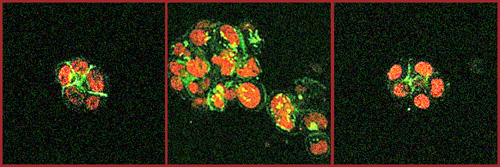
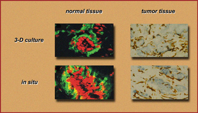
Since she first proposed it in 1982, her ECM theory has been shown to hold true in tissue culture and in mice. These experimental results have not only yielded valuable knowledge about both normal and cancerous breast cells but have also helped reinforce Bissell's fundamental principle that what transpires inside a living cell is greatly influenced by its surrounding microenvironment. This, she says, could explain why not every woman who carries a BRCA 1 or BRCA 2 genetic mutation develops cancer in every one of her breast cells, if she develops cancer at all. It could also explain why such a mutation only causes cancer of the breast or ovaries and not some other form of cancer, like cancer of the skin. In her talk at the Era of Hope meeting, Bissell described how manipulation of the cellular microenvironment can allow malignant cancer cells to revert to a normal appearance and behavior, or phenotype. She also showed how in a tissue culture the microenvironment can determine how a malignant cell responds to therapeutic agents, independently of the characteristics of the malignancy itself. "The ability of breast cancer cells to respond to a therapeutic agent is modified by the microenvironment," she said. "Therapeutic agents can affect breast cancer-cell signaling directly or indirectly, via effects on the microenvironment." Cancer in 3-DBissell has demonstrated that when cancerous breast cells are embedded in 3-D cultures that mimic the architecture of the tissue in their natural setting, they can be reverted to a normal phenotype through the inhibition of various pathways by which the microenvironment sends signals to the cell's genome.
The critical role of the microenvironment's tissue structure was indicated by the fact that these results could not be replicated when the cells were placed in 2-D tissue cultures. The dominance of phenotype over genotype has tremendous implications for cancer therapy, Bissell explained. "Cell growth and malignant behavior are regulated at the level of tissue organization in three dimensions. If the tissue structure in the microenvironment is the 'message,' then tumor cells with abnormal genomes should be capable of becoming phenotypically normal if that structure is restored," she said. "Furthermore, destruction of the microenvironment tissue structure by itself could be a carcinogenic event." As one of the first breast-cancer researchers to make extensive use of 3-D cultures, having used them in experiments for the past three decades, Bissell reiterated the crucial need for such cultures as models for the study of breast cancer and other human diseases.
"The number of drug candidates far exceeds the number of patients, fortunately, in whom one can test them, even if the drugs were found initially to be effective in mice. Also, the cost is prohibitive, even if there were enough patients," she said. "An enormous potential exists in the use of 3-D cell culture models as surrogates for tissues." Bissell has won numerous awards for her research, including the Ernest Orlando Lawrence Award from the U.S. Department of Energy, and the Discovery Health Channel Medical Honor. Her message to attendees of the 2005 Era of Hope meeting, and to breast cancer researchers everywhere, is that there are times when a cell's microenvironment tells the nucleus of the cell what to do and that therefore a new paradigm may be needed to explain how tissue-specific genes are regulated in vivo. Bissell dedicated her 2005 Era of Hope talk to the memory of Sonia Maria Mueller, a Berkeley Lab employee who succumbed to breast cancer after a courageous fight at the age of 35. The series on breast cancer research will continue in the next issue of Science@Berkeley Lab. Additional information
|
|||||||||||||||||||||||||||||||||||||||||
| Top | |||||||||||||||||||||||||||||||||||||||||