|
September 24, 1999 |
|
 |
 |
|
|
|
|
BERKELEY, CA — Using
the Macromolecular Crystallography Facility (MCF) at the Advanced Light
Source (ALS), a team of researchers from the University of California at
Santa Cruz and the Department of Energy's Lawrence Berkeley National
Laboratory has produced the first high-resolution images of a complete
ribosome complex.
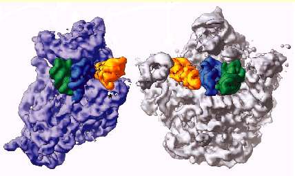
Ribosomes synthesize proteins in the cell by using genetic information to assemble amino acids. The new crystallographic images reveal more about ribosomal structure than any previous observations had suggested, an achievement featured on the cover of this week's issue of Science magazine (Sept. 24, 1999). Harry Noller, a professor of molecular biology at UC Santa Cruz, led the research team, which used the ALS at Berkeley Lab to image the crystal structure of the 70S ribosome of the bacterium Thermus thermophilus at a resolution of 7.8 angstroms (an angstrom is a ten billionth of a meter). Although smaller than most viruses, a bacterial ribosome is a very large molecular complex, consisting of three RNA molecules and more than 50 protein molecules, with a mass of some 2.5 million daltons. "Obtaining atomic-resolution diffraction data for so large a macromolecular complex can only be done with a high-brightness source of x-rays," says Thomas Earnest, a member of the team who is a biophysicist in Berkeley Lab’s Physical Biosciences Division. Earnest is the group leader of the Macromolecular Crystallography Facility. "Beamline 5.0.2, with its combination of flux (the number of photons) and collimation (parallel alignment of the photons), is one of the best in the world for this work."
Ribosomes are tiny organelles responsible for protein synthesis in the cell. They receive genetic information from messenger RNA molecules, which are copies of the gene sequence, and use this to specify the assembly of amino acids, brought by transfer RNA. Until the advent of synchrotron radiation facilities like the ALS, obtaining detailed information about macromolecular structures such as ribosomes was an almost insurmountable challenge. Yet this information is essential for understanding the mechanism by which these critical organelles function. While other research groups have obtained high-resolution images of individual ribosome subunits, Noller and his group, with Earnest, are the first to produce a detailed look at an entire ribosome complex. "My favorite analogy is the cave man coming across a Ferrari," says Noller, referring to the greater understanding that comes with images produced at higher resolution. "There is the moment of discovering the ignition key, the gas pedal, the brakes and steering wheel, followed by years of speculation as to how they work. Eventually, someone lifts the hood, and a new era of experimentation and speculation ensues. Finally, they take the engine, transmission, etc. apart. At this point, there’s a lot more stuff than anyone had anticipated, but at last they have a chance to figure the whole thing out." In addition to Noller and Earnest, the team that created the extraordinary images includes Jamie Cate, now at the Massachusetts Institute of Technology, and Marat Yusupov and Gulnara Yusupova of UC Santa Cruz. From the new 70S ribosome images, Noller says he and the other members of the group are seeing how transfer RNA interacts with the ribosome, and how the two ribosomal subunits interact with each other. In both cases there appear to be complex networks of molecular interactions criss-crossing the ribosome, often involving interactions with a third type of RNA, called ribosomal RNA -- a sort of bridge between the subunits. "One gets the impression that there are systems of long-range communication connecting distant parts of the ribosome," Noller says. "Our images also suggest very strongly that the ribosome is a machine -- and a very complex one with many moving parts. It is also clear that most of the excitement of figuring out the molecular mechanism of translation lies ahead." The MCF at the ALS will be crucial to this effort. The MCF houses three separate beamlines at the ALS's 5.0 complex, all of which are powered by a 38-pole wiggler magnet that provides x-ray photons ranging in wavelength from 0.9 to 4.0 angstroms. The high-end of this energy range, once thought to be beyond the reach of the ALS, is ideal for protein crystallography. A beam of these x-rays sent through a protein crystal creates a diffraction pattern when the photons are scattered by the crystal's atoms. This pattern can be translated by computer into 3-D images of the molecule that makes up the crystal. The announcement of the new ribosome images focuses attention on the status of the MCF as one of the world’s premier facilities for protein crystallography. The demand for time on its beamlines is already heavy: the MCF claims approximately one third of all users of the ALS. To meet an anticipated growth in demand, plans are now underway to add to the ALS three superconducting bend magnets which are capable of generating the higher energy hard x-rays needed for protein crystallography. The addition of these "superbends" would make it possible to boost the MCF’s capabilities to as many as a dozen beamlines by the year 2002. The Berkeley Lab is a U.S. Department of Energy national laboratory located in Berkeley, California. It conducts unclassified scientific research and is managed by the University of California. Additional Information: |