| July 5, 2001 |
|
 |
 |
 |
||
|
|
PEEM2, a second generation photoemission electron microscope, has been operating at the Advanced Light Source for over two years, and, remarks Andreas Scholl, leader of the PEEM2 team, "we have shown that this instrument gets good results" -- which is quite an understatement, says Jun Feng of the ALS. "PEEM2 is the most sophisticated microscope of its type in the world," Feng says, one that has been responsible for a string of benchmark articles in major scientific journals. But while PEEM2 can resolve features separated by just tens of nanometers (billionths of a meter) -- ten times better than the best optical microscopes -- the structures of many complex materials are even smaller. "Characterizing these materials at ultrahigh spatial resolution is essential to scientific and technological progress," says Feng. His hope is to achieve resolution ten times better yet, of one or two nanometers, "the ultimate possible." Feng's team is one of two in the world who are racing to build the next generation PEEM. A collaboration of German universities, research institutions, and corporations called SMART has a head start, but Feng and his colleagues are confident that the experience and resources of their tightly-knit PEEM3 design group, centered at Berkeley Lab, can catch up -- and in the process add some unique features, particularly for the study of magnetic materials. How a PEEM works When a beam of synchrotron x rays is focused on a sample, the material emits electrons whose energy and trajectory convey a great deal of information. PEEMs collect and focus these electrons with a system of magnetic and electrostatic lenses. The resulting image not only gives a physical picture of the sample but can indicate what it's made of and how its atoms and molecules are organized. PEEMs can even look beneath the surface to reveal magnetic structures. Elements and their chemical environments are identified and located by tuning the energy of the incident beam: different energies (frequencies) stimulate electron emission from different elements. If the x rays from the synchrotron source are polarized, PEEM images can reveal the length and orientation of chemical bonds, and -- depending on how the beam is polarized -- depict the orientation of magnetization in magnetic materials. Aberrant behavior
Like an optical microscope, the spatial resolution of a PEEM can be limited by aberration, so-called "image error." Aberration also wastes electrons. Because electrons ejected at the wrong angle or energy can't pass through its focusing system, PEEM2 uses only about five percent of the electrons emitted from a sample. In an optical system like a camera, chromatic aberration produces a rainbow effect when lenses bend light of different colors (wavelengths) at different angles. Spherical aberration produces fuzziness and distortion because a spherical lens or mirror bends light rays at the edges more than at the center. Electrons behave in a similar way: different energies focus at different distances from an electrostatic lens, and electrons focus differently depending on their distance from its center. One way an optical camera can overcome aberration is by correcting the convex lens (which focuses light rays inward) with a concave lens (which spreads light rays apart). But electrostatic lenses only focus inward. To correct electrons, what's needed is a electron mirror. The idea is to send electrons from the sample to a mirror that can exactly compensate for aberrations and then send the electrons back into the optical system. "Essentially we want to reverse the sign of the defect and cancel it," says Feng. Charged reflections
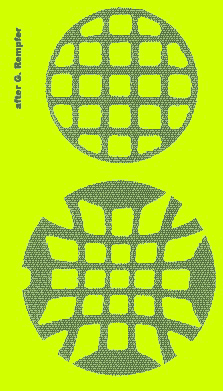
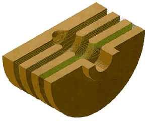
"The most advanced electron mirror so far was demonstrated in 1997 by physicist Gertrude Rempfer at Portland State University," Feng explains. "She proved that a mirror can be used as an aberration corrector, but with her two-electrode mirror, it's very difficult to cover the whole range of aberrations." The PEEM3 team is developing a four-element mirror mounted at right angles to the main column of the microscope; electric fields between the electrodes will cancel aberrations in the beam and reflect electrons back along the path. Design of the mirror and control of the voltages on the electrodes depend on first accurately calculating all aberrations in the electron path. Scholl says, "The voltages can be adjusted within a certain range, but from the viewpoint of the experimenter it's really a tricky proposition, because you have little more than your image to judge from." As a result, the PEEM3 designers are developing computerized schemes to optimize electrode voltages. Among the trickier steps are extracting the aberrant beam and returning the corrected one. An optical system would use a beam-splitter like a prism, but to separate and recombine its electron beam the PEEM3 will use a separator magnet, inside of which the electrons loop around a complicated path and are finally deflected by 90 degrees. "We have several accelerator experts on our design team, including David Robin and Ying Wu, and their expertise in steering and conditioning charged particle beams is invaluable," says Feng. Thanks for your support Crucial to the PEEM3 effort is a synchrotron beam with an extremely high flux, whose energy can be concentrated within the microscope's field of view and whose polarization can be controlled. Beginning this fiscal year, the Department of Energy's Office of Science is funding construction at the ALS of a $3.8 million, elliptically polarized undulator beamline. Critical supporting evidence for the beamline proposal came from the initial design of PEEM3, funded by a Berkeley Lab Laboratory Directed Research and Development Program. Collaborators from many research institutions and industries are participating in the development of the microscope itself. Taiwan's Synchrotron Radiation Research Center (SRRC) -- the first third-generation light source in Asia -- IBM's Almaden Research Center, and the ALS program provide personnel, fabrication facilities, and other contributions in kind; other agencies are expected to fund further development. In addition to Jun Feng, PEEM3 researchers include Simone Anders of IBM; Etienne Forest of Japan's High Energy Accelerator Research Organization, KEK; Howard Padmore, leader of the ALS's Experimental Systems Group; David Robin of Berkeley Lab's Accelerator and Fusion Research Division (AFRD); Michael Scheinfein of Arizona State University; Der-Hsin Wei of SRRC; and Ying Wu of AFRD. Members of Berkeley Lab's Engineering Division include Robert Duarte, Nicholas Kelez, and Ross Schlueter. Andreas Scholl of ALS and Hendrik Ohldag of Stanford work with the team in a consulting capacity.
Additional information:
|