For six decades, this Laboratory has been acknowledged as the birthplace of
nuclear medicine. During Berkeley Lab's 65th anniversary celebration,
pioneering researcher Tom Budinger documented the
Budinger, who heads the Center for Functional Imaging here, chronicled the
preeminent role of the Lab in nuclear medicine -- in the diagnosis and
treatment of diseases, in imaging, and in safety. Berkeley Lab researchers
have provided an ever-clearer window for doctors to view and image disease
within the human body. They have provided physicians new and more effective
ways to treat diseases. And they have devised treatments for diseases that had
been untreatable.
The contributions of nuclear medicine extend to surprising horizons. World
War II aviators, who suffered the bends when flying at high altitudes, were
able to overcome this obstacle thanks to Lab researchers. Radiobiologists here
resolved the mystery of the ghostly flashes of light being observed by spooked
astronauts. Today, researchers are establishing radiation limits for human
space travel.
Radiobiology has further extended these contributions. Melvin Calvin's
Berkeley team resolved the riddle of photosynthesis, discovering the path of
carbon as it travels through a plant, using the tracer carbon-14 (also
discovered at Berkeley). Today, radioisotope tracers are a fundamental tool of
biology.
Budinger, whose Building 50 auditorium audience included many pioneers of
nuclear medicine, started his talk by describing the beginning of the field.
In 1937, Joseph Hamilton was the first to use these tracers to study
circulatory physiology. Using radioactive sodium, Hamilton studied how fast
that which we eat enters and traverses the human body.
Hamilton realized that radioisotopes with a short half-life -- a property
which allows them to be used without medical side effects -- were needed. He
asked the Lab's Glenn Seaborg for help. Seaborg and Jack Livingood bombarded
tellurium with deuterons in the Lab's 37-inch cyclotron, creating iodine-131,
with a half-life of eight days.
Iodine-131 was the beginning of the Lab's ongoing role in the discovery and
use of radioisotopes. In 1938, technetium-99m which remains the most commonly
used isotope in medicine, was discovered by Emilio Segre. Other important
isotopes in which the lab played pivotal roles in the discovery and
application include tritium, carbon-14, fluorine-18 and thallium-201.
During the war, John Lawrence and his colleagues used radioisotopes to help
pilots deal with the consequences of high-altitude flying. Pressurized cabins
did not exist at that point. Donner Lab researchers used radioisotopes of
inert gases to study decompression sickness and other maladies. These tracer
studies made fundamental contributions to the understanding of the circulation
and diffusion of gases. This research led to the development by the
laboratory's Cornelius Tobias of aircraft oxygen measurement equipment. As a
result of this work, an automatic parachute-opener was developed.
Numerous advances were recorded during this era of nuclear medicine at the
laboratory. People suffering from polycythemia vera, a rare disease
characterized by an over-abundance of red blood cells, were treated with doses
of radio-pharmaceuticals. It was the first disease to be controlled with
radioisotopes. In 1940, a pioneering treatment procedure debuted to treat
leukemia. That was also the year in which hyperthyroidism first was diagnosed
and treated using Seaborg and Livingood's iodine-131.
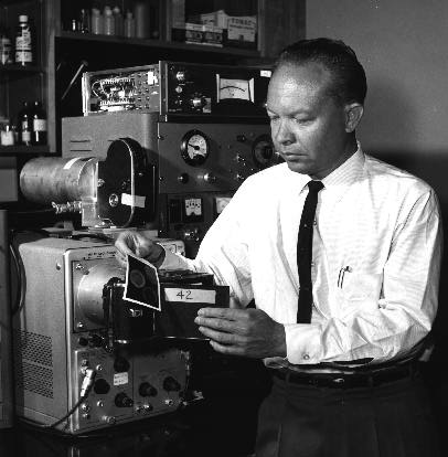
In the 1950s, Hal Anger conducted seminal studies on medical imaging. From
1952 to 1958, he gradually developed the scintillation camera, also known as
the Anger Camera, which enables physicians to detect tumors and conduct other
medical diagnoses by imaging gamma rays emitted by radioactive isotopes.
Developed forty years ago by Anger, these techniques remain the most commonly
used tools in nuclear medicine today.
Many of the applications of the Anger camera and its descendants were
pioneered here. In the 1960s, researchers used the Anger Positron Camera to
diagnose bone tumors. In 1972, Yukio Yano devised a technetium-99m/phosphate
system for bone scanning. In 1979, rubidium-82 was used for dynamic PET
diagnosis of heart disease. Currently, an effort led by Budinger, Derenzo,
Huesman, and Bill Moses is on the verge of creating a 2 millimeter-resolution,
three-dimensional PET camera that can image brain chemistry.
Just as Ernest Lawrence's cyclotrons made possible the creation of
radioisotopes, these accelerators also made possible the use of beams of
neutrons, protons, and heavy ions for the treatment of disease.
In the 1940s here at this Lab, researchers first investigated the use of
neutron beams for cancer radiotherapy. Here in the 1950s, helium and protons
beams first began to be used. Later, in the 1980s, medical researchers here
were the first to begin using heavy ion beams to treat cancerous tumors as
well as a deadly brain disorder known as AVMs, or arteriovenous malformations
(AVMs). AVM is a disease characterized by abnormal growths in the brain of
blood vessels. Heavy ion beams can be precisely focused to obliterate these
growths which, unless treated, can cause lethal or disabling brain hemorrhages
and seizures.
Charged particle beams generated by Lawrence's accelerators have vital
medical uses.
At the Bevalac, which closed in 1993, scores of patients benefited from the
pioneering experimental use of heavy ions to treat cancerous tumors. The
success of this program is responsible for the recent opening of the charged
particle patient treatment facility in Chiba, Japan. This commercial facility
uses a Berkeley Lab accelerator design. A second commercial charged particle
facility is scheduled to start patient treatments in Darmstadt, Germany this
year.
Much of the book on radiation safety was written here.
The Lab's Will Siri literally wrote the first textbook on the safe
application of radioisotopes in biology and medicine. From 1945 to 1979,
researchers developed and refined a model of the effects of inhaled
radioactive particulates. Researchers here have been instrumental in
promulgating guidelines that define the radiation limits of space travel.
These findings have important implications for future interplanetary space
travel by humans.
Budinger is among those whose work is part of the ongoing history of
nuclear medicine. The very day of his talk, Laboratory Director Charles Shank,
Life Sciences Division Director Mina Bissell, and Director of DOE's Office of
Energy Research Martha Krebs joined to praise Budinger at the dedication of
the Lab's new Biomedical Isotope Facility.
Shank said the new facility, which can produce the short-lived
radioisotopes indispensable in many areas of research, would not have been
possible without a stubborn 10-year-effort by Budinger and his chemist
colleagues Chet Mathis and Henry Van Brocklin. As with the nuclear medicine
program begun by John Lawrence, over time, the payoff from persistence and
vision can be of historic dimensions.
extent to which Berkeley Lab has remained the cradle of invention in this
field, right on up to the present moment.

John Lawrence, the father of nuclear medicine
Ernest Lawrence, inventor of the cyclotron, recognized the possibilities for
medicine, and persuaded his brother John to join the Laboratory. John Lawrence
started Donner Laboratory circa 1936. Treating a patient with leukemia, he
administered a radioactive isotope of phosphate. It was the first time that a
radioactive isotope had been used in the treatment of a human disease as well
as the start of a career-long contribution from John Lawrence. He became known
as the father of nuclear medicine and his laboratory is considered the
birthplace of this field.
Ernest Lawrence about the time he came to the
University of California at Berkeley in 1928.

Over time, Anger's scintillation camera evolved into modern imaging systems
such as PET (positron emission tomography) and SPECT (single photon-emission
computed tomography). The evolution of this technology was shaped by Anger,
his colleagues and successors here. Their contributions include the
multi-crystal whole body scanner (1970), gated heart single gamma tomography
(1974), dynamic, gated PET (1978). Today, there are 160 PET cameras operating
in hospitals, medical and research facilities worldwide. The highest
resolution PET scanner in the world -- the 2.6 millimeter-resolution camera --
was built by Budinger's colleagues Steve Derenzo and Ronald Huesman, and
resides here.
Hal Anger , shown with his invention, the positron
scintillation camera