|
July 6, 2001 |
|
 |
 |
|
|
|
|
BERKELEY, CA — Researchers in the laboratory of Alexander Pines, a member of the Materials Sciences Division of Lawrence Berkeley National Laboratory and a professor of chemistry at the University of California at Berkeley, have recovered high-resolution nuclear magnetic resonance (NMR) spectroscopy data from experimental samples in a grossly nonuniform field. NMR pioneer Pines worked with postdoctoral fellow Carlos Meriles and their colleagues Dimitris Sakellariou, Henrike Heise, and Adam Moulé to develop the technique, which could significantly extend the use of NMR spectroscopy as an analytical tool. Their new "ex situ" method is described in the 6 July issue of the journal Science. Until now, high-resolution spectroscopy could only be done "in situ," by placing the sample inside the bore of a very large stationary magnet that produces a strong, uniform magnetic field. With the new technique it may soon be possible to take an NMR probe to otherwise inaccessible samples in the field and obtain high-resolution information. "What makes NMR useful is that can provide a profile of a sample, a kind of fingerprint," says Carlos Meriles, "in which each component has recognizable features." NMR spectroscopy has been used to study the molecular structure and chemical dynamics of a vast range of compounds, materials, and processes, everything from an organism's metabolic state to the composition of promising new materials. The recognizable features in an NMR spectrum show up as distinct peaks of varying height. Until now, in order to clearly resolve two or more separate peaks, the sample had to be placed in a static, uniform magnetic field. Those nuclei in the sample with magnetic moments align their spins, up or down, with the static field. When the sample is irradiated with a radio-frequency (rf) pulse matching the slight energy difference between the up and down spins, it's as if the nuclei are knocked off balance, precessing (wobbling) on a tilted axis around the field lines. Each species of nucleus has a characteristic wobble rate, information that is reemitted as the nuclei relax and realign with the static field. Because the same kinds of atoms may experience different magnetic environments in the presence of other nearby atoms, their signals can differ and point to different chemical arrangements.
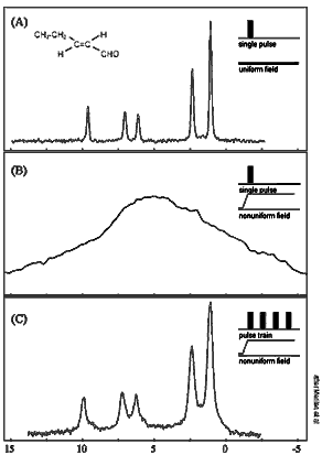
For example, signals from hydrogen nuclei in organic compounds show up as different peaks in an NMR spectrum depending on how the hydrogens are bonded to carbons or other atoms. Displacements from a reference peak are called "chemical shifts" and reflect different concentrations of arrangements of hydrogen in the compound -- producing a spectrum that positively identifies the specific compound. "This way you can tell the difference between, say, oil and water," says Meriles -- just the sort of distinct signatures an oil-well logging instrument looks for in a borehole. In fact, NMR probes have been devised that can be lowered into boreholes, but they necessarily produce uneven magnetic fields, which limits the information they can gather. In a nonuniform magnetic field, slight differences in the magnetic environment of nuclei in one part of a sample are overwhelmed by the much larger difference in static field strength between different locations in the sample. The peaks of different chemical shifts become so broad they blend together, and the spectrum becomes featureless. Once decayed, an NMR signal can be "refocused" by zapping the sample with a second radio-frequency pulse. This produces a so-called Hahn echo, a signal strong enough to detect above the background. But methods based on the Hahn echo erase any spectroscopic information. "The signal still contains information -- it isn't lost, but it's jumbled together," Meriles says. "All you can say is, 'I've got a signal,' not whether it's oil or water, for example." Attempts to get around this roadblock have included analyzing various aspects of the single smeared peak, although "interpretation is extremely qualitative," says Meriles. When Meriles and his colleagues in the Pines lab group set out to solve the problem, they knew one key would be to arrange matters so that the nonuniform static field and the much weaker rf field both fell off smoothly in a correlated way. They did this in the laboratory by imposing a strong spatial variation on the static field, with the rf coil placed at one end of the sample -- thus simulating the conditions of a mobile, ex-situ experiment. To visualize how lost spectroscopic information is recovered, it helps to plot the chemical shift of each spinning nucleus on a three-dimensional graph in which the static field is oriented on the z (up and down) axis, and the rf field, which varies regularly, is thought of as rotating in the x-y plane. Viewed in a frame that rotates at the same frequency, each distinctive chemical shift can be represented by a vector in the x-y plane, rotating around the z axis with that frequency. But in a nonuniform magnetic field, spins get faster or slower, spreading the signal until it overlaps other chemical shifts. The researchers realized that the sharpness of the chemical-shift vectors could be restored if the slow and fast offsets could be exchanged, so that as the signal evolves these components would converge, not spread. To do this they designed a special sequence of pulses of precise energy, duration, and timing. The decaying NMR signal is zapped in such a way that the first rf pulse lifts the vector out of the x-y plane, where it is vulnerable to a second pulse timed to reverse its previous dephasing; a third pulse equal to the first lays the vector back down in the x-y plane but with fast and slow spin segments now reversed. Thus from a decaying signal that might otherwise smear to featurelessness as it evolves in a nonuniform field, the researchers are able to recover and intensify individual chemical shifts to yield a high-resolution NMR spectrum. The Pines group tested their arrangement on a series of compounds, concluding with a liquid known as trans-2-pentenal, whose characteristic spectrum, obtained with a single rf pulse in a uniform static magnetic field, shows the chemical shifts of hydrogen nuclei as five sharp spikes. The same sample, if subjected to a single rf pulse in a nonuniform field (outside the magnet bore), resembles a featureless mound. But if the sample is then subjected to the specially tailored string of pulses in the same nonuniform field, the five peaks are restored to their characteristic positions and amplitudes on the spectrum, with virtually the same sharp resolution. "We have demonstrated that high-resolution NMR spectra can be recovered even with a strongly inhomogeneous magnetic field," says Meriles, "which means it may be possible to develop a mobile magnet that can be scanned over otherwise inaccessible objects to get magnetic resonance information." There is much to be done, he stresses. "You need a really strong field to get a decent decay rate. The stronger the gradient, the worse the problem. It's a challenge to develop a strong magnet that can be taken into the field, or to develop ways to recover a signal from a weak field." But the principle of ex situ NMR spectroscopy has been demonstrated. "Approach to high-resolution ex situ NMR spectroscopy," by Carlos Meriles, Dimitris Sakellariou, Henrike Heise, Adam Moulé, and Alexander Pines, appears in Science, 6 July 2001. The Berkeley Lab is a U.S. Department of Energy national laboratory located in Berkeley, California. It conducts unclassified scientific research and is managed by the University of California.
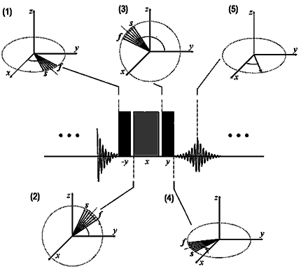
In a nonuniform magnetic
field, the chemical shift of hydrogen, plotted as a vector
in the x-y plane (1) has slow and fast spin components
(marked s and f) which spread the signal. A sequence of
pulses lifts the vector into the y-z plane (2), reverses
its previous dephasing (3), and lays it down again in
the x-y plane (4), with fast and slow components now reversed.
The chemical shift is subsequently recovered and intensified
(5). Additional information:
|