|
December 9, 1999 |
|
 |
 |
|
|
|
|
BERKELEY, CA � A
combination of electron microscopy and single particle image analysis has
been used to produce the first three-dimensional images of the protein
complex that initiates the transcription of DNA's genetic code for the
subsequent production of new proteins. The research was conducted by
scientists with the U.S. Department of Energy's Lawrence Berkeley National
Laboratory (Berkeley Lab) and the University of California at Berkeley.
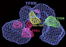
The 3-D images, at a resolution of 35 angstroms (atoms typically have a radius of one to two angstroms), identify critical components of a complex of transcriptional factor (TF) proteins that include TFIID, TFIIA and TFIIB. A model reconstructed from the images shows TFIID as a horseshoe-shaped structure surrounding a central cavity inside of which recognition and binding to DNA takes place. TFIIA and TFIIB are shown to bind to the TFIID in a way that affects the size and shape of the cavity. The research team that produced these images was led by Eva Nogales who holds a joint appointment with Berkeley Lab's Life Sciences Division and UC Berkeley's Molecular and Cell Biology Department. Other team members were Frank Andel, of Berkeley Lab, and Andreas Ladurner, Carla Inouye, and Robert Tjian of UC Berkeley. Tjian is also affiliated with the Howard Hughes Medical Institute. Their results were published in the December 10, 1999 issue of the journal Science. After a strand of DNA has been unwound and unzipped in preparation for protein production, TFIID binds to an exposed DNA section precisely where a genetic message begins. Once TFIID recognizes and binds to a gene on a strand of DNA, it interacts with RNA polymerase so that the genetic code is transcribed into messenger RNA which then carries the information out of the cell's nucleus and into its cytoplasm where proteins are assembled. "Our 3-D reconstruction gives us a good idea as to how TFIID works in concert with TFIIA and TFIIB to initiate and regulate the transcription of protein coding genes," says Nogales.
Structural determinations for some domains and other components of the TFIID, TFIIA and TFIIB proteins have been determined through x-ray crystallography but the size of TFIID, coupled with the difficulties posed in trying to crystallize all of the TF proteins together has so far precluded the use of x-ray diffraction for imaging the entire complex. Consequently, until now, the overall shape and relative position of the components within the complex were a mystery. Imaging via electron microscopy does not require that a protein be crystallized. Because the Berkeley researchers did not have to crystallize the TF complex, they could work with a relatively small amount of sample and still produce a 3-D image of the entire transcriptional machine. In their Science paper, they present TFIID as roughly 200 x 135 x 100 angstroms in size, dominated by three main lobes, each of which measures about 60 angstroms in diameter but differs in structural detail. The lobes are connected by narrow bridges some 20 angstroms wide which are arranged around a cavity whose open channel measures 40 angstroms. This open channel can easily fasten around a single strand of DNA. "The electron microscopy studies presented here reveal a model for the structure of TFIID complexed to both TFIIA and TFIIB in the absence of DNA," the authors write in their paper. "Antibody mapping of the TATA Binding Protein (TBP) within TFIID strongly suggests the binding position of DNA to be at the top of the central cavity within the TFIID complex." To produce the 3-D model, Andel recorded thousands of images of randomly-oriented individual protein molecules in samples obtained from the Tjian research group. A computer was then used to align these thousands of randomly-oriented images into an ordered array and merge them into a three-dimensional reconstruction. In essence, the single particle image analysis was used to create a virtual crystal. "Our study shows that single particle methodology is a useful technique for the structural characterization of mega-Dalton transcription complexes," state the authors in their Science paper. "It also motivates the mapping of individual TBP-associated factors by specific antibodies and higher resolution studies using electron microscopy techniques." At a resolution of 35 angstroms the shapes of TFIID and its companion proteins plus their relative positions within the TF complex can be clearly seen. When this electron microscopy information is combined with the x-ray data on various substructures within the complex -- a technique dubbed "hybrid crystallography" -- Nogales and her colleagues expect to find further clues as to how the transcriptional machinery comes together. Berkeley Lab is a U.S. Department of Energy national laboratory located in Berkeley, California. It conducts unclassified scientific research and is managed by the University of California. Additional Information: |