| April 9, 2004 |
|
 |
 |
| Robots Score Big with Protein Crystallographers | |
| Contact: Lynn Yarris
(510) 486-5375 lcyarris@lbl.gov |
|
| |
BERKELEY, CA � A big step towards speeding up the process of solving protein structures has been achieved by researchers with the Lawrence Berkeley National Laboratory (Berkeley Lab) who have developed and successfully demonstrated the first automated system for mounting and aligning protein crystals in an x-ray beamline at a synchrotron light source. With genome sequencing becoming almost a conveyer-belt process, one of the next big challenges in biology is to determine the structures of the proteins being assembled by all those genomes. In the architecturally loopy, twisted world of proteins, knowing structural form is a key to understanding molecular and cellular function. As there may be more than 30,000 different kinds of human proteins, and nearly a trillion different kinds of proteins on Earth, solving protein structures is another process that screams out for automation.
“Thanks to our automounter robots, in less than three years of operation we’ve been able to screen more than 10,000 protein crystals and collect complete structural datasets on several hundred of them,” says Thomas Earnest, a biophysicist with Berkeley Lab's Physical Biosciences Division who founded the protein crystallography program at the Advanced Light Source (ALS) and who now leads the Structural Proteomics Development Group. Earnest and his group developed the protein crystal automounter robots in collaboration with the bioinstrumentation group of Berkeley Lab’s Engineering Division. The ALS at Berkeley Lab is an electron synchrotron and storage ring, designed to accelerate electrons to energies of nearly 2.0 billion electron volts (GeV) and extract from them – using either bending, wiggler, or undulator magnetic devices – beams of ultraviolet and low energy or "soft" x-ray light. It is home to the Berkeley Center for Structural Biology (BCSB), one of the premier facilities for x-ray protein crystallography in the world today. The BCSB operates eight beamlines, three powered by a multipole wiggler and five by superbends. “Speed is the big advantage of automation,” Earnest says. “Whereas it took about 15 minutes to manually mount and align a crystal, we can now do the same thing in less than three minutes using our automounter robots.” By eliminating the human errors that inevitably set in during a repetitious procedure, and by coupling the robots to a unique software program that enables researchers to cull their best data, Earnest and his colleagues have also been able to improve the quality of the protein crystallography being done at their beamlines. “We were the first to automate the protein crystal mounting process at a synchrotron light source,” Earnest says. “The technology has proven so successful it is now being exported to protein crystallography beamlines at other synchrotron light sources, including the National Synchrotron Light Source at Brookhaven, the Advanced Photon Source at Argonne, and the Cornell High Energy Synchrotron Source.” X-ray protein crystallography is the most widely used method today for determining protein structures. A beam of x-rays is sent through a crystallized protein and scattered by the crystal’s atoms, creating a diffraction pattern of dots whose image can be translated by computer into a 3-D model of the protein. There was a time when x-ray protein crystallography was done with low intensity beams from laboratory x-ray tubes and collecting complete diffraction data sets for a single protein crystal could take months or even years. All this changed with the arrival of synchrotron light sources which can generate x-ray beams that are on the order of a hundred million times more intense than the light from the most powerful x-ray tubes. Now, enough x-ray diffraction data for imaging a typical protein can be collected within an hour, or even, for some proteins, within minutes – providing you can quickly mount and align the protein crystals in the x-ray beamline.
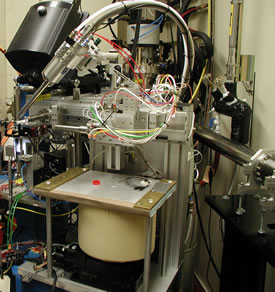
Enter the protein automounter robot systems being used at the ALS crystallography beamlines. Each system features a liquid nitrogen Dewar that can store up to 112 protein crystals, and a cryogenically cooled, moveable arm, called a “gripper,” that plucks crystals from the Dewar and transports them to the beamline goniometer where the diffraction data is collected. The crystals have to be preserved at liquid nitrogen temperatures (about 100 Kelvin) to minimize any radiation damage that might compromise the integrity of the final 3-D structural model. “Our automounter robot system looks like a simple procedure, but having to switch from a liquid nitrogen to an ambient environment creates difficulties,” says Earnest. “One of the biggest challenges is reliability. Because everybody’s crystal is important to them, our robots have to be virtually 100 percent reliable.” As a U.S. Department of Energy national user facility, the ALS and its beamlines are available to qualified researchers throughout the country. To save time for users outside of Berkeley, as well as to further increase their throughput speed, Earnest and his colleagues have also developed special “hockey puck” cassettes, each with an individual capacity of 16 crystal samples. As many as seven can be loaded at once into a standard dry shipping Dewar. Once the robot gripper loads a crystal into the beamline goniometer, unique instrumentation-control software enables users to position the crystal in a way that maximizes diffraction data gathering. A relational database allows users to test and compare crystals for diffraction quality so that only the best crystal from a sample group is used for making a 3-D structural model. The development of this software is a collaboration between Earnest’s group, primarily software engineers John Taylor and Carl Cork, and the Computational Crystallography Group led by Paul Adams, a computer scientist with the Physical Biosciences Division and the Deputy Principal Investigator of the Berkeley Structural Genomics Center. Adams and his team are now working on a software package called PHENIX (Python-based Hierarchical ENvironment for Integrated Xtallography) that will also automate the user software interface for biomolecular structure determination starting with the processed data generate by the beamlines. “Incorporating artificial intelligence into data collection, processing and analysis is the next step,” says Earnest. “With the automounter robot hardware, we’ve given this system a good set of arms and legs, now we want to give it a higher-level brain. Ideally, the process should be like an assembly line with crystals coming in, structures going out.” The first automounter robot system was installed at ALS beamline 5.0.1 in February, 2001. The second and third systems were added to beamlines 5.0.2 and 5.0.3 about a year later. Details about these automounter robots appear in the April 7, 2004 issue of the journal Structure. The paper is titled Automated Sample Mounting and Alignment System for Biological Crystallography at a Synchrotron Source. Earnest is the senior author of the paper. The other authors are Carl Cork, Robert Nordmeyer, Earl Cornell, George Meigs, Derek Yegian, Joseph Jaklevic and Jian Jin of Berkeley Lab, plus Raymond Stevens with the Scripps Research Institute in La Jolla, California, and Gyorgy Snell with Syrrx, Inc., in San Diego. Berkeley Lab is a U.S. Department of Energy national laboratory located in Berkeley, California. It conducts unclassified scientific research and is managed by the University of California. Visit our Website at www.lbl.gov/. Additional Information
|
||||||||||||||
| Top | |||||||||||||||