| lab a-z index | phone book |
| October 13, 2006 | news releases | receive our news releases by email | science@berkeley lab |
|
|||
|
|||
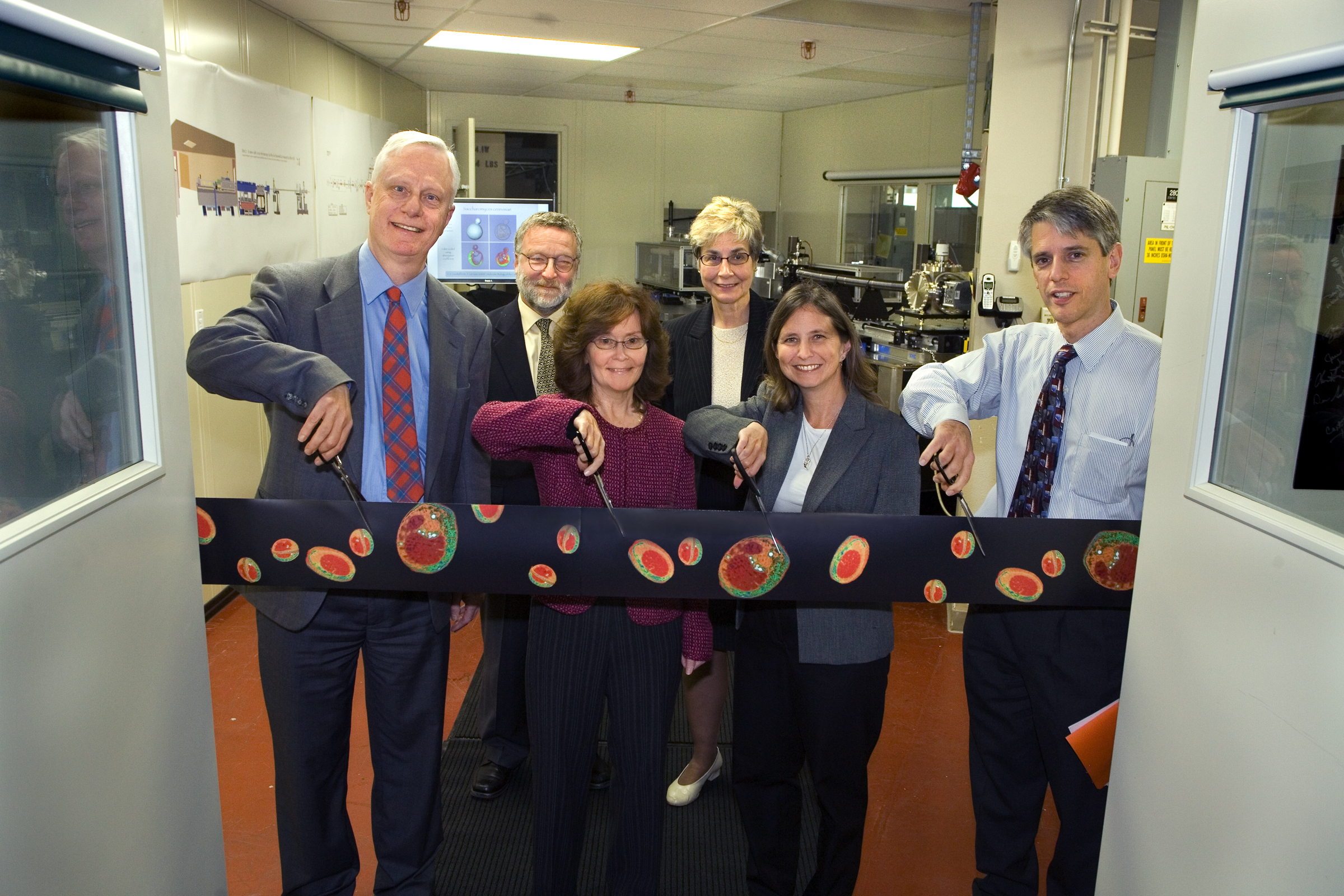
Lawrence Berkeley National Laboratory Opens the National Center for X-ray Tomography |
||||||||||||||||
| Contact: Lynn Yarris (510) 486-5375, lcyarris@lbl.gov | ||||||||||||||||
BERKELEY, CA — The National Center for X-ray Tomography (NCXT) has officially been dedicated at the U.S. Department of Energy’s Lawrence Berkeley National Laboratory (Berkeley Lab). Located at Berkeley Lab’s Advanced Light Source (ALS), this new center features a first-of-its-kind x-ray microscope that will enable scientists to perform “CAT scans” on biological cells, just one of many unprecedented capabilities for cell and molecular biology studies.
“X-ray microscopy is an emerging new technology that expands the imaging toolbox for cell and molecular biologists, and we are going to make this technology available to the greater biological community,” said cell biologist and microscopy expert Carolyn Larabell, who is the principal investigator for the new center. Larabell, holds a joint appointment with Berkeley Lab’s Physical Biosciences Division and the Anatomy Department of UC San Francisco. Her co-principal investigator at the center is The NCXT is being funded with grants from the U.S. Department of Energy (DOE) and from the National Institutes of Health (NIH). For construction and five years of operation, the Biological and Environmental Research (BER) program in DOE’s Office of Science has provided about $7 million. NIH has provided about $5 million through its National Center for Research Resources program, which establishes “biomedical technology resource centers,” such as the NCXT, to provide scientists and clinical researchers with “the environments and tools they need to understand, detect, treat, and prevent a wide range of diseases.” As an NIH technology resource center, the NCXT will be available to qualified biomedical researchers throughout the nation. Said Larabell, “Although there are currently many powerful techniques for imaging biological cells, each with its own unique strengths and limitations, there remains a gap between the information that can be obtained with light microscopy and electron microscopy. X-ray microscopy can bridge this gap by combining some of the best features associated with light and electron microscopy, plus bringing in some entirely new capabilities.”
The centerpiece of the NCXT is the first soft x-ray transmission microscope to be designed specifically for biological and biomedical applications. It is capable of imaging whole, hydrated cells at resolutions of about 35 nanometers, and specific structural elements within the cell at a resolution of at least 25 nanometers. Future improvements could put the resolution of this microscope as fine as 10 nanometers. This superior resolution is made possible by a combination of the high brightness of x-ray light produced off an ALS bend magnet beamline and nanofabricated zone plate optics produced by Berkeley Lab’s Center for X-ray Optics. Berkeley Lab’s ALS is an electron-based synchrotron/storage ring capable of generating x-ray beams that are one hundred million times brighter than those from the best x-ray tubes. The intensity of the beamline will make it possible for users of the NXCT to collect a complete data set for an x-ray tomography image in a matter of minutes, compared to the days and even weeks required for electron microscopy. “With the NCXT microscope, we can examine whole cells, identify subcellular components and locate macromolecules inside cells at substantially better resolutions than light microscopy and without the elaborate specimen preparations needed for electron microscopy,” said Larabell. Microscopy using photons that fall within the visible light region of the electromagnetic spectrum remains the workhorse of biology because it enables scientists to examine living cells in their natural state. Resolution, however, is typically no better than 200 nanometers, and that is only when the light is focused on a single spot. Microscopy techniques based on the use of electrons can provide images at a resolution of five nanometers or better. However, samples must be sliced thin and put through an elaborate chemical preparation and stained in order for the electrons to penetrate and yield high-res images. With transmission x-ray microscopy, samples are rapidly frozen and need no further chemical alteration or staining to be imaged. Because of the quick turnaround time between sample preparation and data collection, scientists using the NCXT will be able to accumulate a statistically significant volume of data within a relatively short time. With transmission x-ray microscopy, samples are rapidly frozen and need no further chemical alteration or staining to be imaged. Furthermore, with the use of metal tags, Special features of the NCXT microscope include improved zone plates, the optic devices composed of nanometer-scale concentric metal rings that are used to focus x-rays for imaging purposes, and greater energy tunability, which means it can be used to obtain images of thicker samples as well as high-res images of thinner samples just by spinning to a different zone plate. Through multiple labeling of proteins, the NCXT microscope can also be used to study protein complexes as well as individual proteins. “In many instances the role played by a protein is determined by its cellular location,” said Larabell. “Soft x-ray tomography is an excellent method for obtaining information on the spatial location and distribution of individual proteins in the cell.” Berkeley Lab is a U.S. Department of Energy national laboratory located in Berkeley, California. It conducts unclassified scientific research and is managed by the University of California. Additional Information
|
||||||||||||||||
| Top | ||||||||||||||||