| lab a-z index | phone book |
| October 23, 2006 | news releases | receive our news releases by email | science@berkeley lab |
|
|||
|
|||
Berkeley Lab Gets $13 Million in Grants from HHMI and NIH to Speed Crystal Structure Solutions |
||||||||||||||||
| Contact: Lynn Yarris (510) 486-5375, lcyarris@lbl.gov | ||||||||||||||||
BERKELEY, CA — With genome sequencing having reached nearly a mass-production mode and new genes being identified on a regular basis, there is a growing demand for faster methods of identifying the structures of the proteins and nucleic acids being produced by these genes. Two new grants to researchers at the U.S. Department of Energy’s Lawrence Berkeley National Laboratory (Berkeley Lab) are aimed at helping to meet this demand by further automating the crystallographic process. The Howard Hughes Medical Institute (HHMI) has awarded $4.8 million to upgrade the robotic capabilities of their crystallography beamlines at Berkeley Lab’s Advanced Light Source, and the National Institutes of Health (NIH) has awarded an $8.2 million grant to further develop a software program called PHENIX, which automates crystallography data acquisition and analysis.
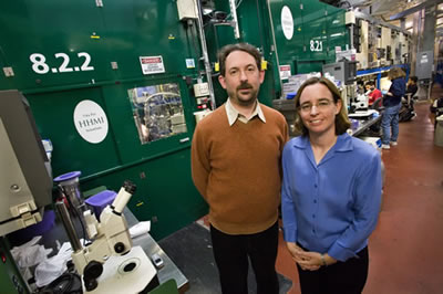
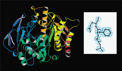
“More and more scientists have come to realize that automating macromolecular crystallography leads to better results,” said Paul Adams, an authority on crystallography who heads the Berkeley Center for Structural Biology (BCSB). “In the time it takes to screen one or two crystals by hand, automation enables 10 or 20 crystals to be screened. The ability to screen many samples prior to data collection enables researchers to focus their studies on the very best samples.” Under Adams’ leadership, the BCSB operates five macromolecular crystallography beamlines at the ALS, including beamlines 8.2.1 and 8.2.2, two “superbend” beamlines which were built through funding by HHMI. The key to understanding a molecule’s function is to determine its three-dimensional structure. X-ray crystallography — using a synchrotron light source like the ALS — is one of the main techniques for accomplishing this. In x-ray crystallography, a beam of x-rays sent through a crystal creates a set of diffraction patterns that can be translated by computer into 3-D images with atomic-scale resolution. ALS beamlines 8.2.1 and 8.2.2 feed off superconducting bend magnets that generate the high-energy "hard" x-rays which are ideal for crystallography. The HHMI is a philanthropy that has become one of the world's foremost biomedical research organizations. Through HHMI’s latest grant to the ALS, beamlines 8.2.1 and 8.2.2 will be equipped with robotic crystal auto-mounting equipment in their experimental end stations that will enable researchers to take advantage of high throughput crystal screening. Beamline 8.2.2 will receive an upgrade of its optics to decrease the size of its x-ray beam from 100 x 150 microns to approximately 30 x 100 microns. This reduction in size will increase the beam’s brightness and permit diffraction experiments to be carried out on crystals as tiny as 10 microns in diameter, about the size of a human cell. Both beamlines will also receive new and improved x-ray fluorescence detectors that will permit the detection of weaker signals from small or dilute samples, and both will also receive upgraded computer hardware. In addition, the end station at beamline 8.2.1 will be provided with an upgraded large-format CCD detector, similar to the one already available on beamline 8.2.2, which will facilitate high-resolution data collection and the study of crystals with large unit cells. “Crystallographic structure solution for a wealth of proteins and other macromolecules has often been hindered by the small size of available crystals,” said Adams. “The ability to focus a high-brightness x-ray beam into a size small enough to match the dimensions of a crystal should allow us to make optimal scientific use of that crystal.” The $8.2 million grant from NIH for PHENIX is an unusually large sum for the development of scientific software, but PHENIX is not typical scientific software. PHENIX stands for Python-based Hierarchical Environment for Integrated Xtallography. It is the product of an international collaboration led by Adams, which, in addition to Berkeley Lab, includes researchers from Los Alamos National Laboratory, Cambridge University in the United Kingdom, Texas A&M, and most recently Duke University. “The PHENIX software provides the necessary algorithms for high-throughput protein structure determination by x-ray crystallography,” said Adams. “It is designed to help both novice and expert crystallographers extract as much meaningful information from their data as possible.” Five years in the making and first released in April of 2005, with a subsequent edition released this past July, PHENIX has already drawn more than 1,000 software downloads. Its current version is highly automated and can rapidly arrive at an initial partial model of a structure without significant human intervention, even when provided with data of moderate resolution and quality. This was made possible through the combination of new algorithms, automated model-building and a comprehensive set of crystallographic libraries.
“These crystallographic libraries, algorithms and automation form a framework that we’re using to develop an expanded system that can start with reduced x-ray diffraction data from any experimental source, and automatically solve and complete protein crystal structures, generate a model or set of models consistent with the data, and help the user prepare those models for deposition,” said Adams. “The impact of this new version of PHENIX should extend beyond the realm of structural genomics and allow crystallographers to also solve other challenging biological problems.” Under the grant from NIH’s National Institute for General Medical Sciences, Adams and his collaborators will develop algorithms for protein structure model completion, the identification of problematic data, and automated decision-making. They will also develop the tools needed to extend the capabilities of PHENIX to nucleic acids. “DNA and RNA represent a very important class of molecule crucial to understanding biology, yet even though more than 1,000 structures containing nucleic acids are in the Worldwide Protein Data Bank, there are no automated procedures for building models of these molecules,” said Adams. “We will fill this gap by developing methods to automatically build, refine and validate nucleic acid structures.” The next stage of development for PHENIX is scheduled to take about five years. The upgrade of ALS beamlines 8.2.1 and 8.2.2 is already underway and is expected to be completed in 2008. Berkeley Lab is a U.S. Department of Energy national laboratory located in Berkeley, California. It conducts unclassified scientific research and is managed by the University of California. Visit our Website at www.lbl.gov. Additional Information |
||||||||||||||||
| Top | ||||||||||||||||