|
Combine three Nobel laureates with one of
the world's brightest x-ray sources, and you're bound to get something big
— or, in this case, exceedingly small. Using Berkeley Lab's Advanced
Light Source (ALS), a highly decorated team of University of Texas Southwestern
Medical Center and Howard Hughes Medical Institute researchers determined
the three-dimensional structure of a protein that controls cholesterol level
in the bloodstream.
Knowing the structure of the protein, a cellular receptor that ensnares
cholesterol-laden low-density lipoprotein (LDL), will help researchers
understand how LDL receptor mutations promote dangerously high cholesterol
levels in some people. And this knowledge could someday lead to treatments
that target these diseases. But it all starts with the protein's structure,
and this starts with a process in which the protein is crystallized and
then analyzed using light rays brighter than the sun.
|
|
 |
|
|
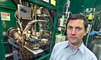
| Keith Henderson helped a team of Nobel laureates
unravel the structure of the LDL receptor protein at ALS beamline
8.2.1. |
|
|
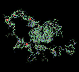
"We were able to see the structure of the LDL receptor's extracellular
domain, and from this we can deduce its function," said Keith Henderson,
a physicist in Berkeley Lab's Physical Biosciences division who helped
the research team select a protein crystal most likely to yield its structural
secrets.
How do you go from crystal to structure to function? The ALS, like all
synchrotrons, accelerates electrons to nearly the speed of light and bends
them in a circular path by powerful magnets. At this speed, electrons
emit extremely bright x-ray light that is directed along a beamline toward
an object that researchers want to investigate at the atomic level, such
as a crystallized protein.
The pattern in which x-rays diffract off of the crystal reveals the protein's
molecular structure, which reveals its function. In this case, the ALS
helped determine precisely how the LDL receptor captures cholesterol,
pulls it inside a cell where it is released and metabolized, and then
cycles back to the cell surface to grab more cholesterol.
Such success requires painstaking preparation. First, the LDL receptor
must be crystallized in just the right way, so it best diffracts x-rays.
This involves a process called crystal screening, in which a slew of promising
protein crystals are evaluated to determine which has the best diffraction
characteristics. In this research, the University of Texas Southwestern
Medical Center team sent Henderson 60 crystals, which he analyzed using
a beamline that can be tuned to resonate at several frequencies.
This important step enables the beamline to be trained on an element
that is specially introduced into each protein crystal. This element,
a so-called anomalous scatterer, allows the collection of several data
sets, each representing a unique diffraction pattern obtained as the beamline
is tuned to various wavelengths around the element's x-ray absorption
edge. These data sets are combined to produce an electron density map
of the crystal that offers enough detail to piece together the protein's
structure. It's an elegant way to image complex proteins, provided the
element is successfully embedded into the crystal — which is where
Henderson's work comes in.
"Out of the 60 crystals, we found a clear candidate, meaning we
knew the anomalous scatterer had stuck and its diffraction quality was
promising," Henderson said.
Then it was back to the lab, where the team spent more than a year refining
the crystal's properties, carefully zeroing in on a specimen good enough
to reveal its molecular framework under x-ray diffraction analysis.
Finally, once the crystal was perfected, the team determined the LDL
receptor's structure using data collected during the commissioning of
ALS beamline 8.2.1. This beamline, part of the sector 8 superbend beamlines
funded by the Howard Hughes Medical Institute, is powered by a superconducting
magnet that produces photons which are nearly twice as bright, at 1 angstrom,
as photons produced by standard bend magnets. Because the average length
of a protein bond is about 1.5 angstroms, or 1.5 hundred-millionths of
a centimeter, these super-bright photons are excellent sources of x-rays
capable of probing the intricacies of protein molecules.
Together, the carefully selected protein crystal and
the state-of-the-art beamline yielded a complete data set that was poured
into powerful computer reconstruction programs. The result is the first
image of the LDL receptor in three dimensions.
"It's further confirmation that the Advanced Light Source is a world-class
facility," Henderson said, adding that the six-year study also lends
credence to time-consuming research that isn't guaranteed to pan out.
"It's high-risk, high-reward science."
The payoff is a better understanding of the molecular breakdowns that
lead to high cholesterol. Normally, the portion of the LDL receptor that
protrudes from a cell wall, called the extracellular domain, binds with
LDL in the liver, pulls its cholesterol cargo inside the cell, and metabolizes
it to replenish hormones, the cell membrane, and vitamin D. But in some
people the receptor's extracellular domain is somehow hobbled, allowing
cholesterol to accumulate in the bloodstream and contributing to life-threatening
diseases such as atherosclerosis.
A common cause of this breakdown is familial hypercholesterolemia, a
hereditary disease that affects about one in 500 people. Researchers have
found more than 900 LDL receptor mutations in people with the disease,
but they didn't know how these mutations disrupt the receptor's function.
Now, with the blueprint of the LDL receptor in hand, they understand how
mutations lead to structural changes, and how structural changes lead
to high cholesterol.
"Without the receptor's structure, we're left with only biochemical
evidence that something is wrong — high cholesterol caused by a
mutation — but we're not sure how," Henderson said. "We're
now using the receptor's form to learn its function."
And with this knowledge, researchers can pursue a pharmaceutical remedy
to familial hypercholesterolemia. So far, the image has solved a longstanding
mystery concerning how the LDL receptor releases cholesterol inside the
cell. Here's how it works: when the LDL receptor's extracellular domain
grabs LDL, it pinches off from the cell surface and sinks inside the cell
to form a sac-like vesicle called an endosome. Next, the endosome becomes
more acidic, which triggers the receptor to discard the LDL. Once free
of LDL, the receptor migrates back to the cell surface — a back-and-forth
journey it repeats many times in its bid to cleanse the bloodstream of
cholesterol.
The image also adds to the illustrious careers of three University of
Texas Southwestern Medical Center researchers. Senior author Johann Deisenhofer,
who is also a Howard Hughes Medical Institute investigator, received the
1988 Nobel Prize in chemistry for research using x-ray crystallography
to reveal the three-dimensional structure of protein in cell membranes.
And in 1985, Michael Brown and Joseph Goldstein shared the Nobel Prize
in physiology or medicine for discoveries concerning cholesterol metabolism.
The study, "Structure of the LDL receptor extracellular domain at
endosomal pH," appeared in the December 20, 2002, issue of Science.
|