 |
|
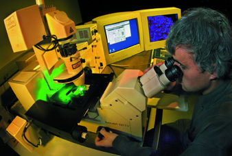
| Stephen Lockett at the confocal microscope in the Life Sciences Division's Bioimaging and Microscopy laboratory. 3-D images of whole cell nuclei can be constructed using this microscope. | |
A mutation in one of the proteins that checks the accuracy of replicated dna may result in daughter cells with aberrant copies of the genome, copies with parts lacking, added, or altered-even with whole chromosomes broken, fused, duplicated, or missing altogether. These cells exhibit a different pattern of protein expression and thus function abnormally; when they divide, abnormalities will be propagated and new ones may be introduced, until eventually cancerous cells result.
"To test this hypothesis in breast cancer development, we needed to analyze the composition of the dna in the individual cells of the tissue at different stages of thedisease," says Lockett. "We needed to detect specific dna sequences in individual cells, and we needed to work with whole cells so we could accurately count the copies of specific dna sequences in each cell. Finally, we had to work with intact tissue, so we could analyze genetic variation from cell to cell."
But techniques developed for standard thin-tissue microscopy preparations, only four micrometers thick, couldn't do the job. Cells and even nuclei are thicker than four micrometers, and standard sections slice right through them, leaving only fragments. In sections 20 micrometers thick or more, however, a majority of cells remain intact. To detect specific dna sequences in thick sections, Lockett and his colleagues use a modification of "fluorescence in situ hybridization" (fish). The modification, developed by Koei Chin in the Cancer Genetics Program led by Joe W. Gray at the University of California at San Francisco, allows dna probe molecules to penetrate thick sections without significantly damaging tissue.
"We stain all the dna in the nuclei using fluorescent probes," Lockett explains. "In our current work we're also labeling specific sites in the center of chromosome one and on the long arm of chromosome 20, a region commonly amplified in breast cancer."
Detecting the labeled elements in thick specimens requires 3-D confocal microscopy. A laser beam, focused through the microscope's objective lens, scans across the specimen at a given depth; the same lens returns the reflected light and the fluorescent glow excited by the laser. While the reflected light is diverted by a dichroic mirror, the faint fluorescent light passes through this beam splitter to a photomultiplier tube. A pinhole mask in front of the photomultiplier eliminates fuzzy, out-of-focus spill.
 |
 |
|
 |
||
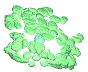
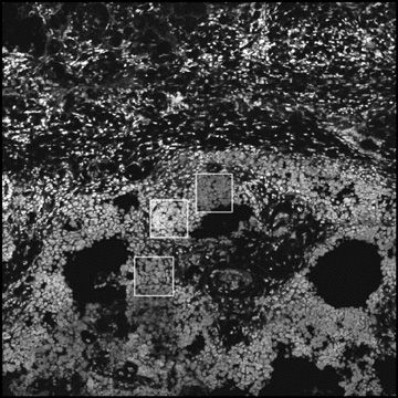
| In the magnified breast tissue above, cancer cells appear lighter. From confocal microscopy of small regions (boxes) the daVinci program constructs images of individual cell nuclei like those at very top; specific genes are labeled with fluorescent probes. | ||
"Since fluorescent stains don't absorb much of the laser light," says Lockett, "a nucleus near the top of a tissue section doesn't shadow the nuclei beneath." Differently stained features fluoresce under different wavelengths of light; whole nuclei glow softly in one color, and the tagged chromosomal sites glow brightly in two other colors.
To distinguish individual nuclei and determine the number of copies of labeled sequences in each, group member Carlos Ortiz de Solórzano, working with colleagues Arthur Jones and Damir Sudar and under a contract with microscope manufacturer Carl Zeiss, Inc., created the "daVinci" program ("data visualization and computer interaction"). A stack of confocal-microscope scans at various depths in the sample is melded into a single 3-D image; the program selects the nuclei, measures their size and shape, and shades them for perspective. The results can be rotated and otherwise manipulated on the computer screen.
To insure daVinci's accuracy, a human expert decides when a candidate is actually a nucleus, a chunk of debris, or a cluster of nuclei which must be further subdivided. "In medical imaging, the de facto gold standard is what you see with your eyes," Lockett says. "Perfect computer visualization programs would have to duplicate the eye-brain system, and we're not quite there yet."
Even the trained human eye may not be sufficient to pick out every doubtful nucleus in a tissue specimen. Divisions in clusters aren't always visible, stains aren't always even, and some objects simply can't be identified. The farther the specimen departs from normal tissue, the harder the task becomes. Experts using daVinci can accurately identify 95 percent of normal human skin cells, 94 percent of cells in benign breast tumors, 89 percent of human breast cells grown in mice (xenografts), and 66 percent of invasive carcinoma cells.
"We continue to strive to improve on this performance," Lockett says, "so we're working with Alessandro Sarti and Ravi Malladi of Computing Sciences on algorithms that can 'denoise' the images." Still, the number of nuclei that daVinci can confidently identify is already enough for measures of genetic diversity.
 |
|
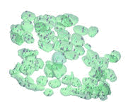
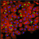
| Above, the computer has counted the number of copies of the gene in each nucleus: green means two copies, the norm, while yellow or blue-only one gene copy or more than two copies-indicates a cancerous cell. | |
"All you have to do is count the number of fluorescent spots in each nucleus," says Ortiz de Solórzano. "In a normal cell there will be two spots marking the centers of chromosome one, and two differently colored spots marking the ends of the long arms of chromosome 20. Any fewer spots or extra spots, and you have an abnormal cell. The more abnormal cells there are in a tissue sample-and the greater the cell-to-cell variation-the higher the level of genetic instability."
Currently, no more than three fluorescent labels can be applied to a thick tissue specimen; fluorescent probes penetrate only a short way, and confocal microscopes can see only a small volume. Lockett and his colleagues have overcome these limitations to long-range experiments by devising ways to acquire and register images of adjacent sections, which can be labeled differently. The new tools and techniques have made it possible to study the mechanisms by which early-stage cancerous lesions spread in the breast.
The Lockett group's collaboration with Gray at ucsf has already shown many genetic changes in the cells of breast cancer specimens, including cell-to-cell variation in genetic properties, a possible hallmark of cancer aggressiveness. They have also shown that tumors may not arise from a single aberrant cell.
"The ability to accurately distinguish nuclei in tissue has general benefits," says Lockett. The model of breast cancer developed by Mina Bissell, director of the Life Sciences Division, shows that the accurate biochemical and physical communication between cells and the complex of proteins in their extracellular matrix (the immediate microenvironment) is crucial to the character and health of cells, and communication breakdown plays a crucial role in triggering cancerous growth.
David Knowles is a group member especially intrigued by the little-explored role of physical structures inside the cell. "Inside, the cell contains organelles that perform numerous functions-mechanisms for protein sorting and tracking, routes for protein travel, structural scaffolding, and so on. How does this cytoskeleton work? How is it connected? How strong is it?"
Working with Mohandas Narla in the Life Sciences Division's Department of Subcellular Structure, Knowles has investigated intracellular structures in red blood cells. "They're good models because they have no nuclei," says Knowles, "but now I'm moving on to 'real' cells."
Using the breast-cancer cell model described above, Knowles, Sophie Lelievre, a researcher in Mina Bissell's laboratory, and William Chou and other members of the Lockett group are investigating changes in the structural proteins of individual cell nuclei that can induce cells to proliferate or, alternately, to arrest growth and differentiate. Through a wide range of studies like these, drawing on the work of numerous collaborators with varied specialties, the members of Lockett's bioimaging laboratory have advanced the hope that underlying
 |
| < Research Review | Top ^ | Next > |