Unraveling
Pseudoknots
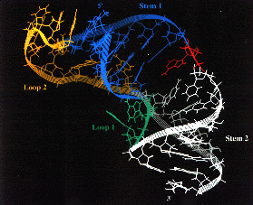
 LBL chemists in the Structural Biology Division, Ignacio
Tinoco, Jr. and Ling Shen, have produced the first three-dimensional image of
an RNA structure that plays a vital role in enabling retroviruses to replicate
within cells. The structure, a double looped strand of RNA that forms what is
called a "pseudoknot," was revealed to contain a bend in its shape that may
serve as the site where key host proteins interact.
LBL chemists in the Structural Biology Division, Ignacio
Tinoco, Jr. and Ling Shen, have produced the first three-dimensional image of
an RNA structure that plays a vital role in enabling retroviruses to replicate
within cells. The structure, a double looped strand of RNA that forms what is
called a "pseudoknot," was revealed to contain a bend in its shape that may
serve as the site where key host proteins interact.
A retrovirus is a protein-coated packet of RNA that requires the chemicals of
a host cell to make
DNA from its RNA genome. When a retrovirus -- the most notorious of which is
HIV -- invades a cell it synthesizes three enzymes (integrase, protease, and
reverse transcriptase) that enable it to transform the host into a virus
replication factory. The mechanism by which this enzyme synthesis is carried
out is called "ribosomal frame-shifting" and involves a shift in the order in
which the virus' RNA genetic code is read. Retroviruses all use a "minus-one"
frameshift, which means the reading of the code starts one nucleotide from
where it should. This controlled frameshifting enables the retrovirus to pack
extra information into its genome.
To understand how pseudoknots promote
frameshifting, scientists need detailed structural information. Tinoco and
Shen, working in collaboration with the UC San Francisco group of current NIH
head Harold Varmus, used nuclear magnetic resonance (NMR) spectroscopy to
produce a three-dimensional high resolution image of a 34-nucleotide pseudoknot
that is known to cause high-efficiency frameshifting in the mouse mammary tumor
virus. In NMR spectroscopy, atomic nuclei are identified and spatially located
by their characteristic absorbance of radiowaves in a magnetic field. Tinoco
has been using this technique to study RNA and in 1992 produced the first 3-D
image of a stem-loop "hairpin," a common and highly stable RNA structural
element with critical folding and protein-recognition properties. When the
loops of a pair of hairpins are joined by base-pairing the combined structure
becomes a pseudoknot. (The structure is only partially twisted, otherwise it
would form a knot.)
In producing their NMR images, the Tinoco and Varmus
research groups experimented with modifying the nucleotide sequences of their
pseudoknots to determine which resulted in frameshifting and which did not.
They discovered that the presence of the nucleotide adenosine at the junction
where the two stems of the pseudoknot are connected creates a bend in the shape
of the pseudoknot. In this form, the pseudoknot promotes high-efficiency (up to
20-percent) sequence frameshifting. If the adenosine is removed, a pseudoknot
is still formed but no frameshifting occurs. The next step will be to find
which ribosomal proteins recognize this bend and interact with it in order to
frameshift. It may be possible, the researchers say, to design drugs that could
fight retroviruses by binding to the pseudoknot and blocking frameshifting.
-- Lynn Yarris
 Return to Highlights Table of Contents