| October 1, 2001 |
|
 |
 |
 |
||
|
|
To learn how tissues develop and maintain their organization — and especially to learn what goes wrong when cancer strikes — it's essential to study individual cells and their nuclei within tissues. The problem is that in real tissues, and in many cell cultures grown in the laboratory, cells are often tightly clustered; their boundaries and the borders of their nuclei are hard to distinguish.
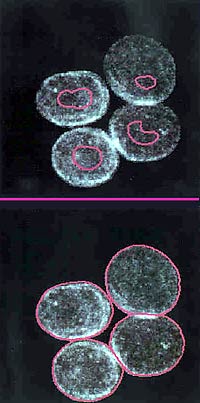
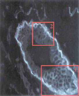
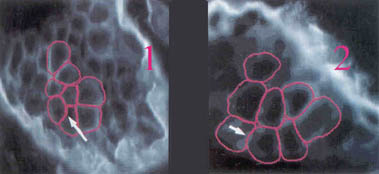
Now Carlos Ortiz de Solorzano of Berkeley Lab's Life Sciences Division and Ravi Malladi of the Lab's Computing Sciences organization, with their colleagues Sophie Leličvre and Stephen Lockett, have joined forces to apply new biological and computational methods to mark and detect the boundaries of closely packed cells and nuclei crowded together under the microscope. Their methods work not only with the envelopes of nuclei but with the membranes of tightly clustered cells as well. To label nuclei the researchers first chose a set of stains that specifically bind to the protein lining of the inner and outer layers of the nuclear membrane, called lamina. Next they developed programs that could find and outline the stained lamina in the microscopic images after they were stored on the computer. "Typically, researchers use fluorescent stains that bind to the DNA inside the nucleus, not to the nuclear envelope," says Ortiz de Solorzano. "But when nuclei are clumped together" — typical in tumor tissue —"there's no contrasting background area between them, and there's no easy way to distinguish one from the next." Seen through the microscope, even stained nuclear membranes may present a mixed bag of dim and cluttered outlines, and while the human eye can pick through these one by one, the new program identifies each nucleus and marks it with a tiny internal graphical "seed," which grows until its periphery corresponds to the outline of the lamina. "Planting just one seed in each nucleus is important, " says Ortiz de Solorzano. "If no seed is planted, the algorithm will be unable to find the boundaries, but if more than one seed is planted, they will mistakenly divide the nucleus into more than one object." Malladi explains that the process begins by automatically choosing seed points to lie inside the nuclei. This is done by computing a crude estimate of the gradient magnitude and direction — a measure of change in the intensity of the image's pixels — in the entire image and translating the gradient points along the "inward" direction. As a result, the gradient points corresponding to the nuclei boundaries reinforce each other in a small region inside nuclei. These ensembles of reinforced gradient points define the initial seed points. "The gradients won't clump exactly in the center," Malladi says, "but that doesn't matter as long as they are inside the nucleus." Next the program moves the periphery of each seed (called the "front") outward until it conforms to the shape defined by the stained lamina. The movement proceeds in stages that are highly sensitive to changes in pixel intensity; the front moves outward freely where changes are minor, but slows almost to a stop when pixel intensity changes markedly, alerting the program to a boundary.
Having marked the inner membrane surface, the front is now allowed to move beyond it, coming to a halt only when it encounters other boundaries expanding from other nuclei. Then the front backtracks, seeking maximum intensity values that clearly identify the stained outer lamina. The algorithm works in similar ways to unambiguously outline whole cells using, for example, fluorescently labeled integrins, which are proteins specific to cell membranes. In both cases "the program makes use of edge-finding algorithms earlier developed for medical imaging," Malladi explains. "We have also developed features that enable the program to find boundaries accurately in very noisy images and to 'see' the right shape even where there are discontinuities." These robust algorithms produce few errors unless the original images are so bad that they are not worth using to begin with.
Computer scientist Malladi is excited about widening the frontiers of software that can abstract visual information from a variety of disparate image sources, ranging from medical images of bones and organs to problems in combustion and fluid mechanics to the underground structures of oil reservoirs. Ortiz de Solorzano, whose focus is biological image analysis, welcomes the nuclei and cell detection techniques as a new means of "quantifying information to understand biological processes." The new techniques are described in the article "Segmentation of nuclei and cells using membrane related protein makers," by C. Ortiz de Solorzano, R. Malladi, S.A. Leličvre, and S.J. Lockett, which appeared in the March, 2001 issue of the Journal of Microscopy. Lockett and Leličvre, formerly of the Life Sciences Division, have recently accepted positions outside Berkeley Lab. Additional information: |