| October 4, 2004 | science beat | | lab a-z index | lab home |
|
|||
| What Lies Beneath (In Buried Nanolayer Interfaces) |
| Contact: Lynn Yarris, lcyarris@lbl.gov |
|
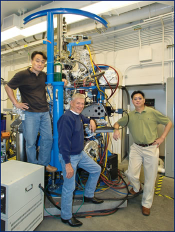
Nanotechnology is superficial, the joke goes, meaning that when objects become so tiny, everything takes place on their surfaces. However, the nanosized components of future computers and memory storage devices will actually consist of multiple overlying layers of material. This means scientists need a way to selectively study the interfaces buried below surfaces. Researchers at the U.S. Department of Energy's Lawrence Berkeley National Laboratory, capitalizing on the exceptionally bright, soft x-ray beams and experimental facilities of the Advanced Light Source (ALS), have developed just such a technique. Charles Fadley, a physicist affiliated with Berkeley Lab's Materials Sciences Division and a professor of physics with the University of California at Davis, led this research effort. His principal collaborators on the project have been See-Hun Yang, now at the IBM Almaden Research Center, and Simon Mun, now an ALS staff scientist.
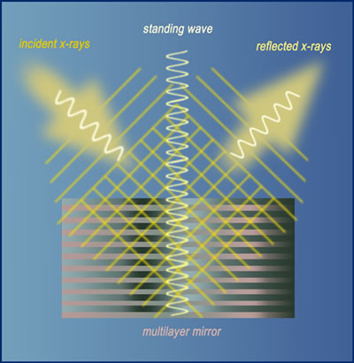
"We've developed a new way of selectively looking below the surface in nanolayers of materials, using soft x-ray standing waves," says Fadley, one of the world's foremost practitioners of the photoelectron spectroscopy technique. "This permits us for the first time to apply all of the principal ALS spectroscopies in a much more depth-sensitive way that directly yields chemical state and magnetic information near the buried interface between two materials." Layered nanostructures will play an especially critical role in the development of the next generation of magnetic read-heads for high-density data storage, and for much-anticipated magnetic random access memory chips, or MRAM. Using spintronics — the spin of electrons rather than their charge — to store data, MRAM promises "instant-on" computers that can store more information and access it faster, while consuming far less power than today's machines. To reach these goals, however, scientists and engineers need a way to "see" what's going on at the all-important interfaces where different layers of materials meet. Furthermore, they need to do this without damaging the materials, a requirement that demands extreme delicacy because each layer is only a few atoms thick. "The most powerful method for studying these interfaces has been scanning transmission electron microscopy, but it requires that samples be sectioned and thinned, so it can't be considered nondestructive," says Fadley. "Using standing waves of soft x-rays, we can nondestructively analyze buried interfaces for atomic composition, and for chemical, magnetic, and electronic structures." A standing wave is a vibrational pattern that is created when two waves of identical frequency interfere with one another while traveling in opposite directions through the same medium. In this case, x-ray waves from the ALS strike a sample grown on top of a nanoscale multilayer mirror, which was designed by researchers at Berkeley Lab's Center for X-Ray Optics to create especially strong reflected waves. When incident ALS x-ray waves interfere with waves reflected from the sample, specific points along the interfering waves appear to be standing still, even though they are vibrating up and down.
"The interfering waves are somewhat like a jump rope, whose ends are fixed but whose midpoint moves up and down," says Fadley. The interactions between standing waves created by the bright, soft x-rays of the ALS and the inner, or core-level, electrons of an atom (as opposed to their outer or valence levels) can be used to identify and study various properties of each atom in the sample. Since Fadley and his research group wanted to investigate the thickness-dependence of the phenomena they were studying, they needed to make their standing-wave probe depth-sensitive. They accomplished this by growing their samples in the shape of a wedge. They then scanned a beam of x-rays across the wedged surface of each sample at an angle that created a vertical standing wave. "The intensity maximum of our standing waves, which generates most of the signal we measure, occurs at a particular depth below the surface," says Fadley. "This gives us the depth-sensitivity and the high resolution we need to map the changes in chemical and magnetic behavior that take place at and around an interface." The beams used to create the standing-wave probe are circularly polarized soft x-rays generated at ALS Beamline 4.0.2. A beam of light is circularly polarized when its electric-field component rotates either clockwise or counterclockwise around the direction in which the beam is traveling. The absorption of circularly polarized light by a magnetic material at an interface reveals much about the magnetic moments of the atoms at that interface. This makes Beamline 4.0.2, which is powered by one of the ALS's premier undulator magnets, ideal for studying the types of buried interfaces that will be found in future data storage devices.
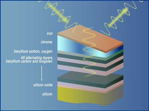
"With soft x-ray standing waves we can, for the first time, look at multilayered nanostructures using a tour de force of all the relevant spectroscopy techniques, including photoelectron, x-ray emission, and x-ray absorption spectroscopies," says Fadley. Fadley and his colleagues have already used their standing-wave technique to gain new insight into the mechanisms behind the phenomenon known as giant magnetoresistance or GMR, a 20 to 50 percent boost in electrical conductivity that occurs when a magnetic field is applied to interfaces between magnetic and nonmagnetic metals. GMR nanostructures can be found in virtually all of today's computer disc read heads, and a detailed understanding of the buried interfaces in these devices is critical to their performance. "We applied the standing-wave technique to an iron/chrome interface and were able to determine the width of the interface, as well as a detailed profile of the magnetic field running through it," Fadley says. "We observed that chrome, which normally is nonmagnetic, was being magnetized by iron just below the interface, but in a direction opposite to the field of the iron." Fadley says that the standing-wave spectroscopy technique he and his group have developed should prove useful for future studies of ultrathin films, liquid layers, molecular clusters, and environmentally related surface chemistry. Combined with an x-ray microscope, it could also be used to do spectroscopy in three dimensions. Additional information
|
| Top |