| June, 2007 | science@berkeley lab | | lab a-z index | lab home |
|
|||
| A Nanoscale Injector for Biological Cells | |||||||||||||||||||||||||
| Contact: Lynn Yarris, lcyarris@lbl.gov | |||||||||||||||||||||||||
The prick of a flu shot may momentarily sting, but the penetration of the needle does no lasting harm to the skin. Likewise, the use of a nanoscale injector to introduce molecules into a biological cell does no harm to the cell.
A team of Berkeley Lab and UC Berkeley scientists have developed such a "nanoinjector" and successfully used it to introduce protein-coated quantum dots into living human cells. The nanoinjector consists of a carbon nanotube attached to the tip of an atomic force microscope (AFM). A special linker molecule connects the designated cargo to the nanotube, which safely delivers it to the inside of a cell within 15 to 30 minutes. "This is the first mechanism capable of delivering cargo into cells with nanometer-scale spatial control and no apparent cellular harm," says chemist Carolyn Bertozzi, an expert in biomimetics who co-led the research with physicist Alex Zettl, a leading authority on carbon nanotubes. Zettl and Bertozzi, an investigator with the Howard Hughes Medical Institute, both hold joint appointments with Berkeley Lab and UC Berkeley. Molecular probes are a critical tool for the study of cell biology. Scientists introduce probes into cells to observe the physical properties and biochemical interactions that govern cellular activities. Among the newest and best optical probes for the study of single particles and single molecules in cellular systems are quantum dots, nanometer-sized crystals of semiconductors like cadmium selenide and cadmium sulfide, which can be engineered to serve as versatile and highly effective fluorescent labels. The trick is to get the quantum dots inside the cell without doing significant structural damage. "The major challenge is to overcome the barrier imposed by the cell's plasma membrane," says Bertozzi. "In the past, this has been accomplished in a variety of ways," including making the membrane permeable using lipids, electric currents, or pore-forming toxins, or by physically penetrating the membrane with a micropipette. "However, all of these techniques result in some physical damage to the cell membrane." Bertozzi and Zettl, working with Xing Chen, a graduate student who is a member of both their research groups, and physics post-doc Andras Kis, set out to develop an alternative method of intracellular delivery that would combine the concept of microinjection with emerging tools from nanotechnology.
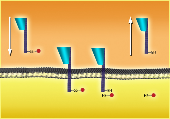
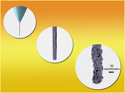
"We envisioned a nanoinjector that would penetrate cell membranes with minimal perturbation, delivering cargo to the cell's interior with high spatial resolution at the nanometer scale," says Bertozzi. "The proposed technology would be comprised of three essential components: a needle with nanoscale diameter, a manipulator with nanoscale resolution, and a controllable means of loading and releasing cargo." In the nanoinjector they devised, a single multiwalled carbon nanotube attached to the tip of an atomic force microscope, or AFM, serves as the nanoneedle. Carbon nanotubes are hollow wires of pure carbon about 50,000 times slimmer than the finest human hair but stronger than steel, making them ideal needles. AFMs, which converge to a point only a few atoms wide, are used primarily to map the surface topography of materials at the atomic scale. When integrated with an inverted fluorescence microscope, the AFM tip becomes an ideal nanomanipulator. For a controllable means of loading and releasing cargo, the Berkeley team used a linker molecule, a disulfide bond between two "bookends," a pyrene section on one side and a biotin section on the other. The pyrene adsorbs to the nanotube, while the biotin binds to the bacterial protein streptavidin, which can be used to coat the surface of the quantum dot. "In the relatively oxidizing environment of the cell's exterior, the disulfide is stable," says Bertozzi. "However, once exposed to the reducing environment of the cytosol" — the internal fluid of the cell — "the disulfide is cleaved, liberating its attached cargo. Since the kinetics of disulfide bond cleavage within mammalian cells has been extensively studied, we can readily predict the rate at which the cargo will be released during the nanoinjection process."
The Berkeley team has already used their nanoinjector to deliver small numbers of streptavidin-coated quantum dots into a line of human cervical epithelial cancer cells to study diffusion kinetics in the cytosol. They were able to deliver a discrete number of molecules to each cell's interior with no need of a carrier solvent and no discernible membrane or cell damage. In the future, they would like to combine the AFM and a confocal fluorescence microscope to carry out organelle-specific nanoinjections. With this capability, they could also introduce probes into plasmid DNA, or carry out parallel nanoinjections of different cell sites with different probes. Additional information
|
|||||||||||||||||||||||||
| Top | |||||||||||||||||||||||||