| August 6, 2007 | science@berkeley lab | | lab a-z index | lab home |
|
|||
| Embedded: A Benign Way to Nanowire Living Cells | |||||||||||||||||||||||||
| Contact: Lynn Yarris, lcyarris@lbl.gov | |||||||||||||||||||||||||
One day it may be possible for physicians to use electrical stimulation to guide the development of embryonic stem cells into neurons, heart cells, lung cells, breast cells, muscles, and other specific cell types. Researchers with Berkeley Lab and UC Berkeley, in collaboration with researchers at the Gladstone Institute of Cardiovascular Disease (GICD) in San Francisco, have taken a critical first step toward that goal.
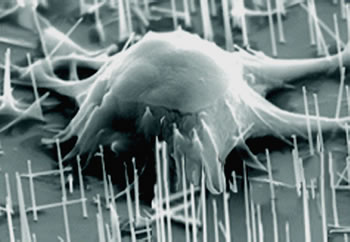
The researchers have developed a technique by which silicon nanowires can be embedded in a living cell, with no apparent harm to the cell. The technique can be used to connect individual cells to one another and to wire the cells to external sensors and other electronic devices. It may also have the potential to deliver genetic material to specific organelles within a cell. "This is the first example of nanowires interfacing with biological cells without the use of external force," says chemist Peidong Yang, who led the research. "The cells were cultured on a silicon substrate that was topped with a vertically aligned silicon nanowire array. The embedding of the silicon nanowire array into individual cells naturally occurred during cell incubation." Yang is a leading nanoscience authority who holds joint appointments with Berkeley Lab's Materials Sciences Division and Molecular Foundry, and with the UC Berkeley Chemistry Department. He worked with Woong Kim and Miki Kunitake of his own research group and with Jennifer Ng and Bruce Conklin of GICD on the embedded-nanowire project; their results are reported in the Journal of the American Chemical Society. Stimulating cell identityDifferentiation is the process by which an embryonic stem cell acquires the unique morphological, genetic, and functional characteristics of a specific type of cell. While controlled by gene expressions, cell differentiation can also be influenced by signals from the surrounding environment, including electrical stimulation. "Whether the signals are physical, chemical, or electrical, the overall effects of these stimuli will eventually control how a stem cell matures," says Yang. "By running an electrical current through a wired stem cell and varying the power through it, we may be able to direct how the cell differentiates." The key to this possibility is being able to embed a living cell with an electrically conducting wire without otherwise impairing the cell's development. Previous attempts to physically puncture cells with nanowires or carbon nanotubes resulted in damage to the cell wall and often the death of the cell itself. Yang and his colleagues avoid this destruction by enabling the cells to gradually incorporate the nanowires by themselves, as they grow and develop. The silicon nanowires were synthesized using a chemical vapor deposition technique developed earlier by Yang and his research group, in which nanosized gold particles serve as a catalyst to trigger the formation of millions of nanowires on the substrate. The precise size of the gold particles controls the diameter of the nanowires. When the silicon nanowires are exposed to air, a layer of silica (silicon dioxide) forms on their surface. "Silica has a proven compatibility with the cell membrane and interior environment, which is one of the reasons we chose to work with silicon nanowires," says Yang. "Also, silicon is a conductor, which opens up the potential for introducing electrical stimulations into the cell." In addition, silicon nanowires have a high aspect ratio — they can be a thousand times longer than they are thick — yet are rigid enough to be mechanically manipulated.
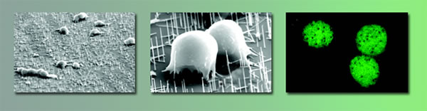
Yang and his colleagues first tested the technique with embryonic stem cells from a mouse. Embryonic stem cells are notoriously sensitive to external stimulation, but when the test cells were grown in solution over the silicon nanowire array, they assimilated the nanowires and continued to thrive for more than a month. The mouse embryonic stem cells used by Yang and his colleagues were on the cusp of differentiating into cardiac muscle cells. Because they had been engineered to express green fluorescent protein (GFP), a combination of confocal and scanning electron microscopy enabled the researchers to observe that each cell had indeed been embedded with several nanowires. "Over time, as these cells proliferated, they began beating like a heart," says Yang. A second round of tests involved human embryonic kidney cells. In these tests, each cell was embedded, on average, with two to three silicon nanowires. The nanowires measured between three and six microns in length, but three different diameters were tested, 30, 90 and 400 nanometers. (A micrometer is a millionth of a meter; a nanometer is a billionth of a meter.) Cells embedded with the 30- and 90-nanometer diameter wires — several orders of magnitude smaller than the cells — survived for up to a week, but those embedded with 400-nanometer wires died within a day. "The longevity of these different cell types has not been systematically compared at this stage, but we can say that it is highly dependent on the diameter and density of the nanowires used in each set of experiments," says Yang. "Also, we need to do further research to understand the mechanism by which the silicon nanowires were taken up by the cells at the molecular level."
Yang and his colleagues also demonstrated the potential of their cell-wiring technique for delivering genes to specific regions of a cell's interior. They coated an array of silicon nanowires with plasmid DNA that codes for the green fluorescent protein. Then they incubated human embryonic kidney cells on top of the wires. One day later, some of the cells began expressing GFP, indicating a successful delivery and normal functioning of the GFP gene. "We believe that the penetration of the nanowires into the cells promotes the retention of the cells on the substrate and therefore the gene delivery," says Yang. "As we perfect this technique, we should be able to pinpoint the delivery of genetic material within a spatial resolution of 50 nanometers, which is far more precise than any existing technique." In addition to applications such as guiding stem cell differentiation and carrying out organelle-specific gene delivery, still down the road, Yang says the cell-wiring technique should quickly become a powerful research tool. "Embedded silicon nanowires could be used as probes of individual cells, or for performing high-resolution, single-cell imaging, or high-resolution chemical and biological extractions," Yang says. Additional information
|
|||||||||||||||||||||||||
| Top | |||||||||||||||||||||||||