Scientists Light Up NMR/MRI of Molecules in Solution

March 28, 1996
Scientists Light Up NMR/MRI of Molecules in Solution |
|
| By Lynn Yarris (LCYarris@LBL.gov) March 28, 1996 |
BERKELEY -- A technique that enhances the sensitivity of nuclear magnetic resonance (NMR) spectroscopy and magnetic resonance imaging (MRI) to molecules in solution offers intriguing new possibilities for chemical research and medical imaging. The technique, which has been reported in the journal Science (March 29), was developed by scientists with the Ernest Orlando Lawrence Berkeley National Laboratory (Berkeley Lab), the University of California at Berkeley, and Tel-Aviv University.
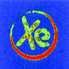
The new technique is based on the use of xenon whose atomic nuclei have been "hyperpolarized" by laser light to give off strong NMR signals. The above images, taken using xenon in solution, hint at the potential of the process.


Xenon is an inert gas that is readily absorbed in solutions and commonly used as an anesthetic. When hyperpolarized xenon gas is bubbled into a solution, polarization from xenon nuclei is transferred to the nuclei of molecules in the solution, amplifying their NMR/MRI signals.
Leading the team of collaborators who developed this new technique were Alexander Pines, with Berkeley Lab's Materials Sciences Division and UCB's Department of Chemistry, and Gil Navon, of the School of Chemistry at Tel-Aviv University. Working with them were Stephan Appelt, Toomas Room, Yi-Qiao Song, and Rebecca Taylor.
Martha Krebs, director of the Office of Energy Research (OER) in the U.S. Department of Energy (DOE) which funded the research, is enthusiastic about the promise of its results.
"The research achievments of Alex Pines have been consistently outstanding for over two decades. These new results are a major step forward in dramatically extending the power of NMR in chemical and biological research."
Both NMR spectroscopy and MRI imaging are based on the tiny magnetic moments produced by the spin of atomic nuclei. Whereas NMR spectroscopy yields a spectrum of lines from individual atoms that can be used to identify molecules, MRI produces recognizable spatial images.
A handicap for NMR/MRI has been the technology's inherent low sensitivity. Obtaining a spectrum or an image depends upon an excess of nuclei in a sample with spins oriented in either an "up" or "down" direction. For an equal number of spins pointing up and down, no NMR/MRI signal would be obtained.
"The natural population difference between up and down nuclear spins is usually no more than one in 100,000 in NMR magnets at room temperature," explains Pines. "This low spin polarization is a persistent challenge for NMR."
To get around that challenge, researchers have resorted to a number of imaginative schemes in which the spins of atomic nuclei in a sample are polarized through assorted combinations of low temperatures, high-pulsed magnetic fields, and exposure to intense microwave radiation.
Alfred Kastler of the University of Paris won the 1966 Nobel prize in physics for his discovery that circularly polarized light can be used to transfer angular momentum from photons to the spins of atoms. Thomas Carver and then William Happer at Princeton University, further developed the "optical-pumping" process and obtained hyperpolarized helium and xenon gas that yielded strong NMR signals which were recently used to image human lungs.
"We have now devised a method that uses both optical pumping and the Nuclear Overhauser Effect (NOE) to transfer enhanced polarization from hyperpolarized xenon gas to molecules in solution," says Pines. "We call this method Spin-Polarization-Induced NOE or SPINOE (pronounced SPIN-OH-EE)."
The researchers say the process that polarizes solution nuclei when they come into contact with the xenon gas is not very efficient. However, this inefficiency is compensated by the hyperpolarization of the xenon nuclei.
"Our strategy is like that of the boxer who punches infrequently but throws blows mostly in the same direction," Pines says. "Every area in a molecule or sample accessed by the hyperpolarized xenon is going to light up to some extent with an enhanced NRM signal."
In their Science paper, Pines and his colleagues reported that with SPINOE, "it is possible to image not only the hyperpolarized xenon but also the molecular environment in which it is accommodated."
Pines and Navon hesitate to speculate about the potential applications of SPINOE to areas of materials and medical research until further experiments have been done. Collaborations, however, are already underway with other research teams at the Berkeley Lab to explore several possibilities. For example, SPINOE might be used to enhance the NMR signals of proteins in solution, or it might be used to follow the trail of hyperpolarized xenon in blood by proton MRI.
The Berkeley Lab is a DOE national laboratory located in Berkeley, California. It conducts unclassified scientific research and is managed by the University of California.