February 22, 1999 |
|
 |
 |
 |
||
BERKELEY, CA -- The successful use of x-ray
crystallography to help identify a protein's molecular function has been reported by
scientists with the U.S. Department of Energy's Lawrence Berkeley National Laboratory
(Berkeley Lab). This demonstration gives science a broad new avenue for interpreting
genetic data.
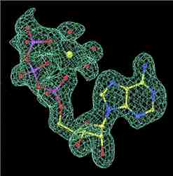
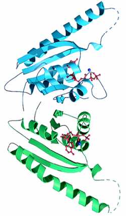
Sung-Hou Kim, a chemist with Berkeley Lab's Physical Biosciences Division and a professor of chemistry with the University of California at Berkeley, led a group of researchers that used intense beams of synchrotron x-rays to determine the three-dimensional crystal structure of a protein whose function was unknown. The protein came from a hyperthermophile, a primitive organism that lives in the hot springs of deep-sea vents. Its structure was found to contain ATP (adenosine triphosphate). The presence of an ATP molecule suggested that the protein functions as either an energy generator, a signal relay, or a molecular switch for activating or de-activating certain chemical reactions. Subsequent biochemical assays indicated this protein serves as a switch. "X-ray crystallography can give us the 3-D structure of a protein from which we can often predict the protein's molecular function (its biochemical and biophysical roles)," says Kim. "These structural predictions provide us with a good lead towards identifying the protein's cellular function, how it networks with other proteins."
This approach to predicting molecular functions based on the structure of a genetically-encoded protein is called "structural genomics," and these results, says Kim, show that it is a viable technique for finding out what a specific gene does. Various projects now underway to decipher the entire complement of genes of several organisms, including the Human Genome Project, promise to revolutionize the fields of medicine and biology in the next century. For any given genome, however, once its full complement of genes has been identified, the critical next step is to determine the one or more molecular and cellular functions of the protein that each of those genes encodes for. "Analysis of several completed genomes shows that many of the encoded proteins cannot be assigned to particular functions, which means that no experimental assays can be easily devised to investigate their exact roles," says Kim. "In other words, scientists have no idea as to what these genes do." The prevailing method for determining the purpose of a specific gene is to compare the sequence pattern of its DNA to that of genes whose roles have already been identified. A major drawback to this approach is that while proteins in different organisms may have similar form and function (the two go hand-in-hand for proteins), the DNA sequencing patterns of their genes may be dramatically different. Given the close relationship between form and function in proteins, solving the structures of proteins to predict their function and classify them into families or groups has posed an attractive alternative. However, until this latest research from Kim and his colleagues, it had not been demonstrated that the structural approach to functional genomics could be done in a timely manner. Essential to his group's success, Kim says, was the use of Berkeley Lab's Advanced Light Source (ALS), a synchrotron that produces beams of x-rays and ultraviolet light for scientific research. Working at the Macromolecular Crystallography Facility, one of the world's premier x-ray beamlines for protein crystallography, Kim and his colleagues were able to resolve the structure of their hyperthermophile protein to within 1.7 angstroms. This high degree of detail not only revealed the presence of a bound ATP molecule unambiguously, but also a new ATP-binding motif that is apparently shared by a family of proteins. "It is estimated that the protein world could be represented by a few thousand families (based on folding domains)," says Kim. "Our results show that through the power of synchrotron radiation, structural genomics is one method by which families of protein structures with hitherto unknown folding patterns could also be discovered." To identify the 3-D structure of the MJ0577 protein from the hyperthermophile Methanococcus jannaschii, Kim and his colleagues first had to crystallize it. One of the reasons this particular protein was chosen as a test-case is that it readily forms a stable crystal. The other reason is that MJ0577 comes from a "deeply rooted organism," one that lies close to the origins of life on the evolutionary scale. "Proteins such as these are often architecturally similar and have the same functions in humans as in bacteria," explains Kim. When a beam of ALS x-rays was sent through the protein's crystal, the incoming photons were scattered by the crystal's atoms, creating a diffraction pattern which Kim and his colleagues were able to translate into a 3-D image. The structure of this protein was then compared against those in the Protein Data Bank at Brookhaven National Laboratory with the idea that any similarities could be used to predict the new protein's molecular properties. Kim's research group has been selected as one of the "test-beds" for a joint structural genomics initiative between the U.S. Department of Energy and the National Institutes of Health. The results of their research on the MJ0577 protein were reported in the December 22, 1998 issue of the Proceedings of the National Academy of Sciences. Members of Kim's research group who contributed to the PNAS paper include Thomas Zarembinski, Li-Wei Hung, Hans-Joachim Mueller-Dieckmann,, Kyeong-Kyu Kim, Hisao Yokota, and Rosalind Kim. Berkeley Lab is a U.S. Department of Energy national laboratory located in Berkeley, California. It conducts unclassified scientific research and is managed by the University of California. Additional information: |