|
July 16th, 2002
|
|
 |
 |
| The BioSig system: finding new meaning in microscopy: Part 2 | ||
| Contact: Paul Preuss, paul_preuss@lbl.gov | ||
|
|
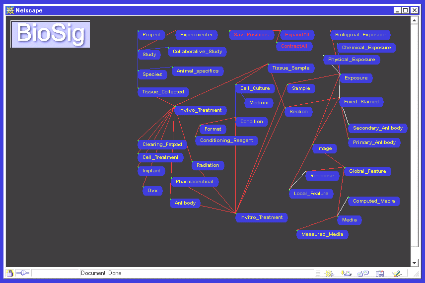
Degrees of freedom "For example, one radiation study compared the wild strain of mice with a genetically altered strain," Parvin says. "Images were collected from mammary sections of mice after they were sacrificed, at intervals of an hour after exposure, four hours after, and eight hours after. How do you compose a picture of time-varying quantitative information, within and across species, for many experimental variables?" The answer Parvin and Barcellos-Hoff came up with was to annotate each image with a wide range of factors, many of which come straight out of the experimenter's lab notebook and can be displayed in a separate window on the BioSig screen. This aspect of the work required the computer science team to gain a detailed understanding of the experimental protocol, a process that involved several sessions with different scientists in the Life Sciences Division.
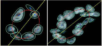
"You want to represent query information visually in terms of plots or images," says Parvin. "But requirements are always changing and new information needs to be added. So the questions facing us were, how do we accommodate such changes in the future? How do we visualize a query result in a way that is informative?" One helpful approach, he remarks, was that "we leveraged the latest dot-com innovations." "One of the best things is, you don't have to redo an experiment to ask new questions," says Barcellos-Hoff. "If you start off looking for one kind of feature in BioSig, but some other feature looks more interesting, you can query the database for just that. For the first time you can follow many different threads through the tapestry." Putting BioSig to work In in vitro experiments, Barcellos-Hoff and her colleagues observed the formation of hollow spheres called acina (Latin for "berries"), a process starting with a single cell in a sheet of human cells of the kind that form the lining of ducts in the breast. The researchers used BioSig to correlate numerous images showing the locations of key regulatory proteins as the acina evolved over a period of 10 days. When challenged by radiation alone, or a protein modifying factor alone, the location of one of the key proteins was disturbed. When cell cultures were subjected to both challenges, however, results were dramatic -- the cells failed to express either of the key proteins, and the acina colonies lost their symmetry, seeming to collapse into their hollow centers.
When what's fuzzy wasn't Barcellos-Hoff agrees. "When I spoke of a fuzzy image, Bahram translated that into a particular distribution of pixels. Sometimes we had to reiterate three or four times to get our concepts lined up." Other kinds of learning were involved as well. "As a microscopist, I was trained to eliminate information, to ignore everything that didn't relate to what I was looking for," says Barcellos-Hoff. "That's a tremendous waste of information -- the opposite of what we're doing with BioSig." But, says Parvin, "this is how you break new ground. One aspect of progress comes from combining multiple disciplines." The ability to save and access vast amounts of quantitative information in images makes them useful in ways never before practical, for example by lending statistical significance to population evaluations. It also constitutes what has been called a "hypothesis-generating data model" -- the potential for testing new ideas by querying experimental information in an existing database, and even importing "legacy" data, which may have been gathered for a completely different purpose. These are among the reasons BioSig research has long been supported by the Office of Biological and Environmental Research in the Department of Energy's Office of Science. In addition, all of BioSig's algorithm developments have been funded through the office of Berkeley Lab's Director. Results are described in detail in the article "BioSig: an imaging bioinformatic system for studying phenomics," by B. Parvin, Q. Yang, G. Fontenay, and M.H. Barcellos-Hoff, in the July, 2002, issue of Computer, published by the Institute of Electrical and Electronic Engineers (IEEE). Additional information: The BioSig system: finding new meaning in microscopy, Part 1 |
||||||||||||||||