|
September 27, 2002
|
|
 |
 |
| Death and resurrection in 3-D | ||
| Contact: Paul Preuss, paul_preuss@lbl.gov | ||
|
|
Most cells in higher organisms know when it's time to die, for the good of the whole multicellular being. But tumor cells infamously resist death, whether from chemotherapeutic drugs or the body's own immune system; finding out how is a major goal of medical and biological research. A new study in the journal Cancer Cell (September, 2002) reports that the formation of cellular structures like those in the breast confers resistance to cell death in both normal and tumor cells. The study was conducted by Valerie Weaver of the University of Pennsylvania's Department of Pathology and Laboratory Science, a member of that university's Institute for Medicine and Engineering, in collaboration with Mina Bissell of Berkeley Lab's Life Sciences Division and others; the work had its beginning when Weaver was a postdoctoral fellow in Bissell's laboratory. When cells refuse to die Programmed cell death, or apoptosis, is the process by which organisms remodel their tissues, ridding themselves of cells infected by viruses, cells with damaged DNA, and no-longer-needed cells that could become dangerous -- like immune-system killer cells when the threat of infection is past. While much is now known about the regulation of apoptosis by specific genes and proteins within individual cells, tumors remain puzzling. What allows some tumors to evade cell death while others succumb to chemotherapy or other treatment? "Multidrug resistance of tumors is a major problem facing the treatment of cancer," Weaver says, "but the mechanisms of resistance are not clear. Cells can be made resistant in two-dimensional cultures -- if sometimes only to one class of drugs -- but when they're put back into a living body, they lose resistance again." Weaver, Bissell, and their colleagues found that an important part of the puzzle involves the way cell structures are organized in the tissues. Since three-dimensional architectures are lost in two-dimensional cell cultures, the vital role shape plays in cellular function has long been obscure. "Of course researchers understand that tumors grow in 3-D," Weaver says. "But critics of studies of multidrug resistance in vivo say, 'there are so many other things going on in the body, it's not easy to demonstrate the role of structure.'" Bissell emphasizes that "cells need to be studied in the context
of their environment, in which growth factors, hormones, and the extracellular
matrix, or ECM, all play vital parts. When forming tissues, you need the
right kind of cross-talk among all these signalling molecules." A model of living tissue To study resistance to apoptosis, Weaver, Bissell, and their colleagues used an exceptional 3-D model of tissue from cultured human breast cells. The model, initially developed in Bissell's lab for rodents and later extended to human cells in collaboration with Denmark's Ole Petersen, now of the University of Liverpool, is capable of distinguishing between normal and malignant cells. In the 3-D cultured-cell model, unlike experiments in living organisms, variables can be controlled and studied one by one.
In the model, nonmalignant mammary epithelial cells -- epithelial cells are those that form internal and external linings -- form attachments to a form of extracellular matrix called "reconstituted basement membrane." In living organisms the basement membrane, consisting of layers of specialized proteins, is essential to anchor cells in place in the breast and many other organs. "The ECM consists of a mass of huge proteins that are secreted outside
the cell," Bissell says. "As far as normal cells are concerned,
it used to be thought that it was needed just to provide a scaffold. But
it does much more: it communicates powerful signals that affect cell behavior
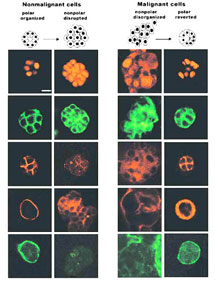
and tissue organization." The 3-D model includes a reconstituted basement membrane, similar to that in a living organism, which induces nonmalignant cells to form hollow spheroids -- "polarized" structures that, as Bissell puts it, "know which way is up." Remarkably, the cells in these spheroids are resistant to apoptosis. By contrast, malignant cells proliferate to form disorganized, nonpolarized aggregates, susceptible to cell death induced by a variety of drugs and other agents used to kill cells. Polar structures of normal cells in the model resemble organoids called acini (Latin for berries) that secrete milk in mammary tissue. Weaver emphasizes that it is not necessary to create working acini in order to confer resistance to cell death: the key is polarity itself. In the model, formerly disorganized cell aggregates can be made to "revert," forming polarized structures through interaction with the reconstituted basement membrane. Once they have formed spheroids, the malignant cells also become resistant to apoptosis. In monolayer cultures, however, both nonmalignant and malignant cells
were equally vulnerable to cell death induced by chemotherapeutic drugs
and immune regulators. The researchers sought other reasons for the stability of cells in polar structures, like their growth status -- how fast the cells were dividing. Unlike what has been believed from clinical studies, however, growth status did not affect resistance to apoptosis. And since the cultured cells were genetically identical, whether in monolayer or 3-D, the results indicated that context can override genetic makeup. How could structure make such a crucial difference? Polar structures
do not arise in a vacuum. The specific molecular and biochemical pathways
by which cells communicate with one another and attach to the basement
membrane not only influence polarity but hold the key to resistance to
apoptosis. |
||||||||||||||||||||