|
August 26, 2002
|
|
 |
 |
| Taking structural studies to a new plateau | ||
| Contact: Paul Preuss, paul_preuss@lbl.gov | ||
|
|
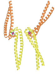
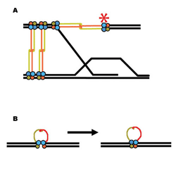
Less than a year ago the National Cancer Institute launched a multidisciplinary "program project" named Structural Cell Biology of DNA Repair Machines (SBDR), aimed at elevating the understanding of DNA repair from the level of individual protein structures to the level of complex interactions within the cell. On August 1, Nature published the project's first major result, the unexpected discovery of the Rad50 "zinc hook," a metal-mediated interface that the essential Mre11/Rad50 protein complex uses to link broken DNA strands so that they can be rejoined.
SBDR is a $18 million program involving 18 investigators at 11 institutions, pursuing five research programs in three major areas of DNA repair. Based at Berkeley Lab, SBDR's principal investigator is John Tainer, a professor of molecular biology at the Scripps Research Institute, with a visiting faculty appointment in Berkeley Lab's Life Sciences Division. Co-principal investigator is Priscilla Cooper, acting director of the Life Sciences Division. "We believe that SBDR will be powerful for providing insights on the function of dynamically assembled DNA repair machines," says Tainer. "These insights absolutely require advanced technologies that bridge the gaps from molecular structure to assemblies to repair networks to cell biology. Such a combination of technologies cannot be found in individual laboratories but instead requires a highly collaborative effort." Cooper adds, "The huge advantages we get from basing the SBDR Program at LBNL come directly from the broad base of biological expertise in DNA damage and repair in the Life Sciences Division, coupled to the structural expertise and advanced biophysical technologies at the Advanced Light Source. The established strength of Berkeley Lab in mounting and sustaining large multidisciplinary programs provides an added major advantage." Understanding complex molecular machines requires an arsenal of techniques, among them traditional molecular biology and biochemistry laboratory methods, electron microscopy, mass spectrometry, and the use of synchrotron radiation. An essential component of SBDR will be the versatile SIBYLS beamline funded by the Department of Energy and now being constructed by the same team who built the Advanced Light Source's highly successful superbend beamlines for structural biology, led by Dave Plate and Nicholas Kelez under the guidance of physicist Alastair MacDowell and ALS Experimental Systems Group leader Howard Padmore. SIBYLS, an acronym for "structurally integrated biology for life sciences," will have the unique capability of performing both x-ray crystallography, to determine the structure of crystallized proteins, and small-angle x-ray scattering (SAXS), which studies proteins in solution, conditions more nearly resembling their natural environment. When the crystal structures of individual proteins are known, SAXS can sometimes be used to reveal how they work together in complexes; it can also be an important tool for gaining structural information on impossible-to-crystallize proteins. Life under siege Different proteins act together along different pathways to repair different kinds of damage; some groups have been doing DNA repair for millions or billions of years and are found in organisms from bacteria to humans. While the specific proteins involved in a particular repair process are not always identical among different organisms, their group geometries and functional "strategies" are often similar.
"Previously DNA repair was thought of as a collection of clean, neat, separate pathways, but now we know that these pathways are interwoven," says Susan Tsutakawa of the Life Sciences Division, a crystallographer in SBDR's molecular biology lab. "Proteins that were thought to have a role in only one pathway are now understood to be involved in many." The proteins Mre11 and Rad50, for example, form repair complexes found in all kingdoms of life; the Mre11 complex's remarkably diverse functions include roles in cell division, maintenance of chromosome-capping telomeres, and repairing double-stranded breaks in DNA in two different ways. The August 1 Nature paper by Tainer and his colleagues compares the Mre11 complex in eight organisms: a virus, an archaeon, a bacterium, two yeasts, a nematode, a plant, and humans. They found a new and surprising mechanism by which the Mre11 complex accomplishes DNA repair. |
||||||||||||||||||||||||