
Frontiers in Nanoscience, page two
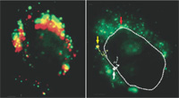
Biologists may soon use nanotechnology to watch the inner workings of a living cell and track the effectiveness of disease-fighting drugs. These two images portray the movement of nanosized probes, called quantum dots, as they pass through a cell's membrane.
In the growing world of nanosized particles, structures and devices, one of the most compelling stories has been that of quantum dots, semiconductor nanocrystals that light up like neon in a rainbow of sharp colors when bathed in ultraviolet light. Qdots, as they are known, have already fueled several startup high-tech companies, including one spun off from Berkeley Lab. Paul Alivisatos, a Berkeley Lab-UC Berkeley chemist who directs the Materials Sciences Division, is Associate Laboratory Director and one of the founders of quantum dot technology. He and his research team recently added an important new chapter to this unfolding story when they combined quantum dots with segmented nanorods to produce an extensive new array of multi-branched nanostructures.
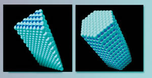
A Lab and UC research team achieved a breakthrough by growing nanowires out of the highly prized semiconductor gallium nitride and then controling the direction in which the nanowires grew. Gallium nitride nanowires are triangular in cross section when grown on a substrate of lithium aluminum oxide but hexagonal when grown on a magnesium oxide substrate. Growth direction is critical to determining a nanowire's electrical and thermal conductivity and other important properties.
Furthermore, they learned to tune the separate components of these nanostructures and calculate the electronic interactions of their branches in three dimensions. This makes it possible to create electronic devices tailored to a variety of applications, ranging from quantum computing to artificial photosynthesis.
In another study, quantum dots were used by a Berkeley Lab and Lawrence Livermore National Laboratory team as nanosized probes for looking inside the nuclei of biological cells. The cell nucleus has been called one of the best known but least understood of all cell organelles, a knowledge gap that stems from the lack of a way to image long-term phenomena within the nuclei. Berkeley’s Fanqing Chen and Livermore’s Daniele Gerion found a way to transport silica-coated quantum dots inside cell nuclei. To slip their quantum dots past the membrane that guards entrance to the nucleus, Chen and Gerion stole a trick from the SV40 virus, which gets through the barrier with the help of a protein that binds to a cell’s nuclear trafficking mechanism. The researchers obtained a portion of this protein and attached it to their quantum dot, creating a hybrid, part biological molecule and part nanosized semiconductor, small enough to slide through the nuclear membrane’s pores, and believable enough to fool its defenses. They’ve been able to introduce and retain quantum dots in nuclei for up to a week without harming the cell. The dots fluoresce for days at sufficient resolution to detect biological events carried out by single molecules. This should allow scientists to track specific chemical reactions inside nuclei, such as how proteins help repair damaged DNA.